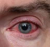
Do people die of basal cell carcinoma?


Do people die of basal cell carcinoma?

OVERVIEW

The most prevalent kind of skin cancer is called basal-cell carcinoma (BCC), sometimes referred to as rodent ulcer, basal-cell cancer, or basalioma. It frequently manifests as a raised, painless patch of skin that may be glossy and has tiny blood veins running across it. It could also show up as an elevated, ulcerated region. Although basal-cell carcinoma can damage the surrounding tissue and grow slowly, it is unlikely to spread to other locations or cause death.

What's BCC?

  • Basal cell carcinoma is curable in sun-exposed areas like the face and neck. 
  • It mostly affects epidermal basal cells. Basal cells are essential for skin regeneration and healing.
  • BCC is called a rodent ulcer due to its sluggish growth and limited spread risk.
  • It seldom extends beyond the skin, unlike melanoma.
  • Sun exposure is the main cause of BCC. 
  • UV radiation from sunshine or tanning beds can cause BCC over time.

Who is more prone to getting basal cell carcinoma (BCC)?

  • Age: BCC is more common in adults over 50. 
  • Fair skin is riskier. 
  • Sun exposure from outdoor activities, sunbathing
  • Employment in the sun.
  • Previous BCC:
  • Significantly more prevalent in men and newborn males.
  • More common in light-eyed people (blue or green eyes).
  • Although BCCs are not considered genetic, certain risk factors might run in families.
  • Fair skin, freckling, and burning rather than tanning are examples.
  • Genetic diseases like Gorlin syndrome can cause BCCs; however, they are rare.

Appearance: 

basal cell carcinom

  • output: BCCs might vary in appearance. They may appear as:
  • Shingles
  • A bleeding, unhealed scab.
  • New red or pearly skin lump.
  • Some have a pearl-like rim surrounding a central crater,
  • Others are superficial and scaly.
  • Untreated, they can create ulcers, hence the name “rodent ulcers.”

Reason for Basal Cell Carcinoma?

  • Sunlight and indoor tanning beds release UV radiation, which destroys DNA in basal cells, the skin's outermost layer. This damage can cause BCCs over time.
  • Genetics affect UV skin response.
  • Age and Skin Type: Later in life, skin gets thinner and less effective at repairing UV damage. The elderly are more at risk.
  • Fair-skinned people, especially those who burn easily and infrequently tan, are more prone to BCCs.
  • Immunosuppression: Medications or conditions that weaken the immune system can raise BCC risk.
  • Skin cancer may occur in organ transplant recipients who take immunosuppressive medicines.
  • Prior Skin Cancer:
  • Having a BCC or other skin cancer raises your risk of getting another.

Basal Cell Carcinoma Signs

  • BCCs tend to be persistent open sores that do not heal adequately. These lesions may bleed, leak, or crust.
  • An open sore may last weeks or heal and reappear.
  • Red, irritated patches:
  • Watch for red spots on your face, chest, shoulder, arm, or leg. Irritated, crusty, itchy, or painful patches may occur.
  • BCCs might look like benign pimples or sores, so don't ignore any unexpected changes.
  • Shiny lumps or nodules: 
  • A flat, waxy region that resembles a scar may indicate BCC. The skin might be white, yellow, or waxy.
  • Scar-like spots may signify invasive BCC.
  • Check for tiny pink growth with a little elevated, rolled edge. The center indentation may be crusty.
  • This growth may eventually create microscopic surface blood vessels.

How is basal cell carcinoma (BCC) detected and treated?

The video about the treatment of basal cell carcinoma


Skin Exam: Your doctor will examine the questionable spot and the rest of your body for lesions.

Skin Biopsy: A skin biopsy can confirm BCC and establish its subtype. This process involves sending a tiny lesion sample to a lab for pathological investigation. BCC type and skin cancer diagnosis are determined by the biopsy.

Treatment Options:

Complete cancer eradication is the main goal of BCC treatment.
Treatment choice relies on numerous factors:
Cancer type, location, and size.
Whether first-time or repeated.
How you follow up and your preferences.

Common treatments are:
Cancerous lesions and healthy skin margins are surgically removed. This approach suits BCCs that rarely reoccur.
Mohs Surgery: Layer-by-layer growth removal with microscopic analysis to assure full eradication. Mohs surgery is advised for facial BCCs at high risk.
Curettage and Electrodesiccation (C and E): Using an electric needle to treat the base of the lesion and scraping the skin cancer's surface

Basal Cell Cancer Treatment
Basal cell carcinoma (BCC) is a common skin cancer that starts in basal cells, which produce new skin cells. Different methods can cure BCC, depending on its nature, location, and health. Explore the 

Primary treatments:
Surgery: Excisional Biopsy: Cancer is surgically removed with a healthy skin margin. Often the first treatment for early-stage BCC.

Cryosurgery: Skin is frozen to generate a scab that slips off. It fits smaller BCCs.
A skin graft from another place of the body may be utilized to cover a significant skin removal.
High-energy radiation is used in radiotherapy to kill cancer cells. When the BCC is widespread, radiotherapy may be considered.

Cancer location
  • Cancer location makes surgery difficult.
  • You're too sick for surgery.
  • Your lymph glands were invaded by cancer.
  • Sometimes radiation is given after surgery to prevent recurrence.
Medicines and Therapy
Also, read https://www.pennstatehealth.org/services-treatments/basal-cell-carcinoma
  • Targeted medicines and immunotherapy try to stop cancer growth, while immunotherapy aids in identifying and eliminating cancer cells.
  • These drugs are available as skin lotions, pills, or IV infusions.
  • They may be utilized if BCC is in many areas, has progressed deeper into the skin, or surgery or radiotherapy isn't possible.
  • The photodynamic therapy technique employs a light-sensitive drug and a light source to specifically eliminate cancer cells.
  • Photodynamic therapy works for thin, non-invasive, non-melanoma skin malignancies.

Post-op scarring

After surgery or injury, scarring is normal. Scars often result during skin healing. Every scar and recovery journey is different. The facts about scars:
  • Scar appearance and development: Initially, your scar may seem reddish. It gradually settles, becoming paler and smoother.
  • The severity of the wound and your healing capacity can delay healing for two years or more.
  • Initially, the stitch line where the incision was created after surgery, especially skin surgery, may seem red.
  • Be prepared for elevated stitches in the first week.
  • A soft pink color gradually replaces the redness over several months.

Scar Care:

  • Scars can't be entirely removed; however, they can be improved.
  • Massage: Aqueous or E45 cream should be gently massaged into your scar several times a day. Only do this once the wound heals.
  • For at least a year, cover your scar in the sun. Protect it with clothing or a dressing.
  • Sunscreen: Apply SPF 30 or higher straight to your scar in the sun.
  • Skin camouflage: Use lotions and powders to hide your scar. They can be tailored to your skin tone and used efficiently.
  • For assistance, visit your GP if your scar is unpleasant or bothersome.
  • Urgently seek medical attention if your scar is swollen, painful, or heated to the touch.
  • Showing infection indications (pus).
  • Scars reveal perseverance and healing. Treat them well, and they'll fade gracefully!

Stages of BCC

Explore basal cell carcinoma phases. These stages assist in guiding treatment and revealing cancer behavior. Remember that BCC is slow-growing and rarely spreads, yet being aware is crucial:

Stage 0 (Carcinoma in Situ): Cancer cells remain contained and have not spread beyond their initial site.
Bowen's illness refers to squamous cell carcinoma in situ.
Consider it a precancerous stage where cells are turning cancerous but haven't infiltrated adjacent tissues.

Stage 1: BCC is classified as Stage 1 if the tumor is 2 cm or less.
The malignancy has not spread beyond the skin.

In Stage 2, BCCs greater than 2 cm but less than 4 cm are still confined but more substantial.

Stage 3 means various things:
The tumor exceeds 4 cm.
It has reached neighboring bones, inflicting slight harm.
It entered nerve space.
It has penetrated the subcutaneous fat layer.
Stage 3 includes tumors less than 4 cm that have progressed to one lymph node on the same side. Importantly, the lymph node's outer layer is intact.

Stage 4 is advanced sickness.
Cancer has spread to lymph nodes.
It has grown through the lymph nodes in the skin.
Multinodal spread.
Spread to opposite-sided lymph nodes.
Stage 4 can also involve distant spread to the bones, lungs, or bone marrow.

Basal cell cancer prevention

Basal cell carcinoma (BCC) is a common skin cancer, and prevention can greatly reduce your risk. The following are some helpful skin-protection pointers:

  • Use sunscreen with an SPF of 30 or higher when you are outside to protect yourself from the sun. 
  • Cover your face, neck, and ears.
  • Find shade from 10 a.m. to 4 p.m. If outdoors, utilize shade or an umbrella.

Safe Clothing: Cover your skin with loose, tightly woven garments. Protect your face and eyes with a wide-brimmed hat and sunglasses.
Avoid Tanning Beds: UV radiation from tanning beds raises skin cancer risk. Get a sun-kissed look with safer options.

Regular Skin Exams:
Check your skin often. Check for new or changing moles, lumps, or lesions.
If you discover a scab that bleeds and doesn't mend, a new red or pearly lump, or a flat, scaly reddish spot, visit a doctor.

Protect Scars and Heal Wounds: BCCs may form in long-standing scars. Protect scars from the sun.
Proper wound care ensures complete healing and reduces problems.

Conclusion

Remember that BCC grows slowly and is usually treatable. Regular skin exams and sun protection are necessary. Consult a dermatologist for skin changes or concerns. Keep using sunscreen to protect against UV radiation! 


How to make blepharitis go away quickly

How to make blepharitis go away quickly

Synopsis

Blepharitis refers to the inflammation that occurs at the corners of the eyelids. In addition to looking oily and crusted with scales that stick to the lashes, the eyelids may become inflamed and uncomfortable. Blepharitis occasionally wakes up with their eyelids glued together. Others can awaken with a sensation of sand in their eyes and dried tears around their eyelids.

blepharitis
Blepharitis

What you should know is as follows:

Blepharitis Types:
  1. Picture the edge of your eyelid if you are experiencing anterior blepharitis. Anterior blepharitis is the term for inflammation of that region.
  2. Pay attention to the inner edge of your eyelid if you have posterior blepharitis. when posterior blepharitis is present in that region. You can occasionally have both kinds at once.

Symptoms: 

  • Having the sensation of a little stone in your eye
  • Frequently blinking
  • Dry eyes
  • watery eyes
  • Redness around the eyelid margins
  • Eyelid swelling
  • The skin surrounding your eyes is flaky.
  • Itching

Reasons:

Blepharitis anterior:
  • Blepharitis caused by Staphylococci: Typically, bacteria reside on your face and eyelashes, but occasionally they overstay.
  • Blepharitis due to seborrhea: Because of Dandruff
  • Additional offenders include viruses, pests, allergies, and cosmetics accidents.
Non-Spreadability and Causes:
  • Blepharitis can't spread to other persons. Therefore, there's no need to send your eyes to eyelid rehabilitation sessions or place them in quarantine.

Another Cause 

  • Bacterial Blockage in Oil Glands:
  • Skin Disorders:
  • The fancy term for flaky, itchy skin is seborrheic dermatitis.
  • Lack of Oil:
  • Rosacea is a condition marked by redness, especially on the nose and cheeks. However, occasionally it also invites the eyelids.
  • The condition known as herpes simplex dermatitis can result in ocular pain and irritation.
  • Seborrheic dermatitis is similar to the more complex form of dandruff.

The skin around the eyes that is flaky and irritated?

  • Dermatitis caused by Staphylococci:
  • The condition is known as allergic dermatitis.
  • Demodex, or demodecosis:
  • Demodex mites are minuscule, small organisms.
  • Infections by parasites:
  • Blockage of the oil glands causes posterior blepharitis.
  • Rosacea (redness of the face)
  • Dandruff on the scalp
  • When your oil glands slow down and your eyes get dry, it's known as meibomian gland dysfunction (MGD).

Risk factors: 

  • Blepharitis can affect anyone.
  • These consist of:
  • Individuals who adore eyelash extensions
  • Individuals who have a history of allergies
  • People who like to massage their eyes and those who wear contact lenses

How may blepharitis be avoided?

Eyelid Hygiene: 
Maintaining cleanliness is essential. As your symptoms become better, you can start cleaning them twice a day and eventually once a day. Think of it as a warm bubble bath for your eyelids but with a clean flannel or cotton wool soaked in warm water instead. After applying it to your closed eyelids for five to ten minutes, give them a gentle massage for thirty seconds.
Crusts and Flakes Be Gone: 
Gently wipe the edge of your eyelashes with cotton wool or a cotton bud. Add a small bit of baby shampoo to water to give your eyelashes a spa treatment if you want to add a little extra luxury.
Before going to bed, take off all eye makeup as part of cosmetics etiquette.
Eyeliner Boundaries:
Steer clear of applying eyeliner on your eyelids' back margins.
Makeup with Contact Lenses: Lens-Free Zone: Give your contact lenses a vacation while you're battling blepharitis.

How Can Blepharitis Be Identified?

1. Eye test:
The eye test station is where we start. The slit-lamp analysis
2. Differential Diagnosis: 
The eye specialist eliminates other possible causes. Is it an impostor or blepharitis? They will take into account ailments such as malignancies of the eyelids or immune-mediated problems that resemble blepharitis.
3. The course of treatment:
To control the unruly eyelids, the ophthalmologist may suggest a topical antibiotic cream or ointment.
Antibiotic eyedrops or tablets may be recommended in more complex cases if blepharitis has resulted in further issues.
Additionally, the ophthalmologist could recommend that you see an eye specialist if the situation becomes too serious.
4. Keep in Mind: Non-Contagious!
It will not spread to other individuals. Consult the professionals if you ever think your eyelashes are organizing a covert party.

Blepharitis Mild Flare-Up Duration:
  1. With appropriate care, mild cases usually begin to get better in a week or so. Severe instances: It may take longer for more serious instances to stabilize. Weeks, or perhaps a few months, are in question.
  2. Chronic Nature: Accept that it's frequently a chronic illness. Although there isn't a permanent cure, the correct care can manage symptoms and lessen them.
  3. Sometimes, symptoms even engage in a game of hide-and-seek, going months or years without showing up before choosing to RSVP once again.

Options for Blepharitis Treatment and Medication

The video explains what you can do with blepharitis.


  • Make sure your eyelids are clean.
  • Warm compresses
  • Using steroid eye drops or antibiotics
  • Also, keep in mind that blepharitis is not communicable.

Pharmacy Expertise:

  • Your neighborhood pharmacist can provide wise counsel. They could suggest:
  • Eye Wipes and Pads: Maintaining Cleanliness and Exclusivity.
  • Eyedrops: Give your eyes a little respite.
  • It's time to hire experts if your eyelids are still problematic after all the TLC.

Your doctor may recommend:

Blepharitis is typically treated with both home remedies and medical care.

Also, read https://www.windsoreyeclinic.com/eye-conditions/blepharitis/
  • In addition to the treatments listed below, a doctor may prescribe the following treatments at home:
  • By using electrochemical lid margin debridement (BlephEx), any bacteria, mites, and the biofilm they produce are eliminated from the eyelids. Additionally, it releases any obstructed Meibomian glands.
  • Any substance blocking the Meibomian glands is melted by thermal pulsation treatment (Lipiflow).
  • Clogged eyelid glands can be opened with intense pulsed light therapy (IPL).
  • In some circumstances, oral or topical antibiotics may be necessary for severe blepharitis.
  • Additionally, your general practitioner may refer you to an ophthalmologist if your eyelids are acting out in a full-blown tantrum. They are the ones with the enigmatic potions and magnified glasses.

Conclusion

  • Poor cleanliness is not the cause of blepharitis.
  • Redness and stickiness of the eyelid, together with clumping of scaly skin around the base of the eyelashes, are the most noticeable symptoms of blepharitis.
  • The goal of treatment is to reduce symptoms; the illness cannot be cured.


Bacterial meningitis can lead to death.

Bacterial meningitis can lead to death.

Overview

Bacterial meningitis can lead to death. Your brain and spinal cord's protective membranes become inflamed when you have meningitis. Inflammation frequently results from brain and spinal cord fluid infections. Meningitis symptoms require rapid medical attention. Deadly bacterial meningitis can kill in hours.

Bacterial Meningitis Can Be Causing Death
Bacterial meningitis

Let's examine essential points:

The symptoms of bacterial meningitis can arise suddenly and may include:

  • High fever
  • Vomiting
  • Headache
  • Not all meningitis patients get a rash. 
  • (that doesn't diminish when rolled over with a glass.)
  • A stiff neck
  • Bright light aversion
  • Sleepiness or inattention
  • Epileptic fits

These symptoms may arise in any sequence, and not everyone has them.

Causes and Transmission: 

  • Both viruses and bacteria can cause meningitis.
  • Although rare, bacterial meningitis is worse than viral meningitis.
  • Kissing, sneezing, and coughing can transmit it.
  • Even healthy people can spread the illness if they have these bacteria in their throat or nose.

Vaccinations:

Certain causes are prevented by meningitis vaccinations:

  • Baby MenB vaccine
  • 6-in-1 baby vaccine
  • Baby and elderly pneumococcal vaccine
  • Baby Hib/MenC vaccination
  • Baby and toddler MMR vaccinations
  • Teens and university students receive MenACWY vaccination.

Who risks meningitis?

Age Groups: Newborns: Newborns are most vulnerable. Bacterial meningitis is especially dangerous for infants under 3 months old. They get bacterial meningitis 70 times more often than adults. Thankfully, routine vaccines safeguard them.

Toddlers: Meningitis and septicemia can still affect young toddlers despite immunizations.

Teens and young adults are at risk of meningococcal illness. These bacteria can live harmlessly in the nose and throat, but teens and young adults are more likely to have them, increasing their disease risk.

Over 65s: Some meningitis forms are more likely to affect them. Older persons are more likely to get pneumococcal illness. Some countries promote pneumococcal vaccinations for seniors.

Environment and Geography: Certain Countries: The prevalence of septicemia and meningitis varies by nation. The incidence is higher in some areas.

Smoke and Mass Gatherings: Tobacco smoke and mass gatherings can increase the risk of meningitis and septicemia. Bacterial meningitis can lead to death.

Also, read https://www.brainandspine.org.uk/health-information/fact-sheets/meningitis/

Medical Issues:

  • Weakened Immune System: HIV/AIDS patients and immunosuppressive drug users are at risk.
  • Although most occurrences of meningitis are isolated, close contact with someone with bacterial meningitis can increase the risk for others.

Treatment for meningitis

The video discusses the treatment of children.


Bacterial meningitis demands immediate hospital care.

  • Directly injected antibiotics and intravenous fluids.
  • Oxygen via mask
  • Viral Meningitis: Mild Cases: Mild viral meningitis usually resolves on its own within 7–10 days. Supportive care is the main treatment.
  • Rest: Rest helps your body heal.
  • Dehydration:  Drink plenty of water.
  • Pain relief: Over-the-counter fever reducers and painkillers can assist.
  • Bacterial meningitis demands hospitalization due to its severity. 
  • Bacterial meningitis can lead to death.

Treatment usually includes:

  • Antibiotics: The bacterial infection is treated with intravenous antibiotics.
  • Bacterial meningitis is life-threatening, thus treatment begins before diagnosis.
  • Intravenous fluids prevent dehydration.
  • Face masks can offer oxygen for breathing problems.
  • Steroids: Some steroids diminish brain swelling.

Duration/Recovery:

  • Patients with bacterial meningitis usually stay in the hospital for a week.
  • Home Recovery: If testing shows moderate viral meningitis is viral, you can go home. Most recover within 7-10 days.
  • At home, practice self-care by resting and relaxing.
  • Treat headaches and aches with medications.
  • Anti-sickness medication helps with nausea and vomiting.

Long-term help:

  • Feeling normal after returning home may take a while. Complications like hearing loss may require additional treatment and care.
  • Preventing Meningitis Spread: The risk of infectious transmission is often low.
  • Family, roommates, and romantic partners of meningitis patients may receive antibiotics.
  • Short encounters with infected people rarely require antibiotics.

How to avoid bacterial meningitis?

Vaccinations:

  • The MenB vaccine is administered to infants at 8 weeks, 16 weeks, and then again at 1 year.
  • Newborns are administered the 6-in-1 vaccine at 8, 12, and 16 weeks.
  • Pneumococcal vaccine: Babies get two shots at 12 weeks and 1 year, and 65-year-olds get one.
  • Babies receive the Hib/MenC vaccine at 1 year.
  • Babies receive the MMR vaccine at one year and again at three and four months.
  • Teens, sixth-formers, and university students can get MenACWY.

Healthy Habits:

  • Give Up Smoking:
  • Get enough rest:
  • Clean Hands:
  • Avoid Sick People:
  • Cover Your Coughs and Sneezes:

Is meningitis serious?

In many cases of bacterial meningitis, prompt treatment can avoid long-term harm. Some may experience brain damage, hearing loss, disability, and seizures for the rest of their lives, even with prompt treatment. Some meningitis patients die.

Home meningitis test?

Push your head forward slowly by pressing on the back of your neck. Hire someone for greater results. Check if your hips and knees flex instinctively when you raise your head. This positive Brudzinski sign suggests meningitis.

Are bacterial meningitis patients totally recovered?

Most meningitis and sepsis patients recover, although a third will have aftereffects. Most aftereffects are short-term or modest, although hearing, brain injury, and amputation can occur.

Do meningitis patients lose hair?

The worst is bacterial meningitis, which can cause permanent brain damage, hair loss, learning impairments, and death. Knowing the symptoms, causes, and treatments for this potentially lethal illness is crucial.

Can meningitis survivors live normally?

Most meningitis patients recover, although others may suffer side effects. Temporary aftereffects may improve. However, some suffer lifelong repercussions.

How to avoid meningitis?

  • Get vaccinated if possible.
  • Use soap and water often.
  • Cover your mouth and nose when coughing or sneezing.
  • Disinfect commonly touched areas.
  • People with contagious diseases should avoid contact.

How long is meningitis?

Bacterial meningitis Hospital stays are normally 1–2 weeks, but complications can extend them. Recovery can take months. Rare yet serious bacterial meningitis affects the brain and spinal cord.

What does the meningitis vaccine cost in India?

Meningococcal immunization costs depend on the vaccine type, brand, manufacturer, taxes, and provider. The cost of each dose of the meningococcal vaccine ranges from Rs 4,000 to Rs 6,000.

Does meningitis surgery exist?

In patients unresponsive to medical therapy, such as failure of drug transport to CSF or pus occlusion of fourth ventricle outlets, traumatic injuries like compound skull fractures or CNS penetrating wounds, and bacterial complications, surgical assistance may be needed.

What deficiency causes meningitis?

Membrane attack complexes cannot form without complement, reducing the immunological response to Neisseria meningitidis. This weakened immune response makes meningitis recurrent.

Foods should be avoided for meningitis.

  • Listeria-containing foods such as soft cheeses.
  • hot dogs.
  • sandwich meats.

What are meningitis stages?

  • Tuberculous meningitis is staged by neurologic status:
  • Stage 1: No mental function changes, impairments, or hydrocephalus.
  • Stage 2: Neurologic impairment and confusion.
  • Third stage: stupor.

What foods aid brain recovery?

  • Brain-feeding protein is important.
  • Traumatic brain injury survivors require more protein to recuperate.
  • Lean turkey, meat, fish, eggs, beans, and lentils are excellent choices. 
  • Adopt Mediterranean eating.

Conclusion

Smoke and Mass Gatherings: Weakened Immune Systems, HIV/AIDS patients, and immunosuppressive drug users are at risk. Bacterial meningitis can lead to death. People can keep watching and avoid the above occurrences, which will prevent this disease to some extent.


How To Get Rid Of Right Lower Abdominal Pain

How To Get Rid Of Right Lower Abdominal Pain

Overview

It's normal to have lower right abdominal pain, and it will go away on its own in a day or two. However, if you're having severe pain or prolonged discomfort, shortness of breath, blood in your vomit, or vaginal bleeding, you should consult a doctor.

Right Lower Abdominal Pain

Let's investigate a few potential reasons:

How can I determine whether my side discomfort is severe?

A closer look is necessary for side pain:

Gas and Constipation: Although they are mostly benign, gases and constipation can cause side aches. Although they rarely represent a significant threat, these troublemakers can cause quite a bit of discomfort. If the discomfort fluctuates, shifts depending on your posture, and goes away in an hour

Irritable Bowel Syndrome (IBS): can cause your large intestine to spasm, resulting in pain, bloating, and cramping.

Gallstones and kidney stones are similar to microscopic geological formations seen inside the body. Your kidneys may have kidney stones, which are composed of minerals and salt, and they can be extremely painful.
The right side is preferred by gallstones, which are made of bile and cholesterol. It is important to seek professional advice if you are feeling radiating discomfort.

Infections: It's like your body is giving you an urgent warning if you have a fever in addition to your sideache.

Sciatica is a type of lower back pain that can radiate down your leg, albeit it is not precisely a side pain. When a nerve becomes irritable, sciatica results.

How can lower right abdomen pain be relieved?

  • For less serious situations, take into account these natural solutions:
  • Rest: Your body may simply want some time to recuperate.
  • Drugs that are sold over the counter: Acetaminophen and ibuprofen are examples of over-the-counter pain medications that may be helpful.
  • Heated Compress: The affected area may feel better after using a heated heating pad.
  • Chamomile Tea: Well-known for its calming effects, chamomile tea can help reduce pain.
  • Gentle Movement: To release tension, try yoga stretches or deep breathing techniques.

What signs of appendicitis are present?

Appendicitis:

  1. Appendicitis is an inflammation of the appendix, a small pouch-like organ that is attached to the large intestine.
  2. Symptoms include an acute lower right abdominal ache that gets worse when you move or breathe. Because an inflamed appendix can rupture, it might need to be treated by a doctor right away.
  3. Take action: If you think you may have appendicitis, get medical attention right away.
  4. The disorder known as appendicitis occurs when the appendix, a long, finger-shaped pouch in the lower right abdomen, swells and becomes infected. Since appendicitis can cause major consequences if left untreated, it is imperative to recognize its signs.

Here are some things to be aware of:

Pain in the abdomen is the defining symptom. Usually, it begins near your belly button before moving to the lower right portion of your abdomen. Sharp, ongoing pain that becomes worse with time is possible. It might be made worse by moving, coughing, or applying pressure to the region. Interestingly, it might be beneficial to raise your knees to your chest.

The video explains lower abdominal pain relief.


  • Vomiting and Nausea: Appendicitis frequently causes nausea and vomiting.
  • Loss of Appetite: Take this as a possible indication if you abruptly lose interest in eating, especially if it is accompanied by stomach pain.
  • Fever: Appendicitis is frequently accompanied by a mild to moderate fever, typically between 99°F and 102°F.
  • Changes in Bowel Habits: You may have diarrhea or constipation. Your digestive tract seems to be performing the cha-cha.
  • Swollen Belly: Inflammation can cause your abdomen to feel swollen or bloated.

How is a diagnosis of appendicitis made?

Physical Examination: The doctor will conduct a physical examination when you see them. To check for pain and edema, they will apply light pressure to your abdomen. It's intriguing to note that the pain will frequently worsen if they abruptly relieve the pressure while your appendicitis is acting up. Your appendix seems to be playing hide-and-seek, but it's not adept at hiding! 

Blood Examinations: A high white blood cell count can be checked with a blood test. An infection or inflammation may be indicated by elevated white blood cells, which is a warning sign for appendicitis.

Urine Test (Urinalysis): Urine tests are occasionally performed to rule out other medical disorders. Although it's a piece of the puzzle, it won't diagnose appendicitis directly.

Imaging Examinations: These are similar to internal detective tools:

Ultrasound It's not only for baby checkups! An ultrasound can help rule out other possible causes of pain, such as kidney stones or ovarian cysts, or confirm appendicitis.

A sophisticated 3D X-ray called a CT scan can identify appendix swelling. It's similar to doing a high-tech picture shoot for your appendix.

Magnetic resonance imaging, or MRI, is helpful when avoiding needless radiation exposure, particularly during pregnancy.

In some situations, doctors might keep you in the hospital for a while to monitor the course of your symptoms. However, without waiting for additional testing, they may suggest surgery (an appendectomy) if it's quite obvious that your appendix is having a fit.

The illusive quest to prevent appendicitis!

  • High-Fiber Diet: Consuming foods high in fiber, such as fruits, whole grains, legumes, and flaxseeds, appears to reduce the incidence of appendicitis.
  • Think of fiber as the express lane for nutrients; it keeps everything going smoothly. It facilitates waste's timely removal from the building.
  • Exercise: You should work out even your appendix! Engaging in regular exercise may lower your risk of developing appendicitis.
  • Vitamin Supplements: According to some research, some vitamins, such as vitamin D, may have protective properties.
  • Maintaining a lofty posture may help prevent appendicitis. Perhaps your appendix likes to stand tall and proud.
  • Fiber Quest: Eat a lot of whole grains, flaxseeds, and legumes, which are high in fiber. Fiber maintains the flow of your intestines, much like a well-rehearsed dance routine.
  • Get those limbs moving with Exercise: Expedition! Your appendix may undergo a small vitamin voyage as a result of regular exercise. Vitamins, not pirate riches, may be your pals. Don't go overboard, though; you don't have to become a human gummy bear.

Can appendicitis be brought on by stress?

An episode of appendicitis is not triggered by stress.
The brain-gut axis is a connection that functions similarly to their covert hotline. That hotline is called when stress strikes.
The Scrolls of Ancient Study: Researchers first looked at the riddle of appendicitis back in 1992. They discovered a tenuous connection between appendicitis and significant stressful events, such as auto accidents or job losses. Your Gut Symphony and Stress:
Irritable bowel syndrome (IBS), the mischievous side effect of appendicitis, IBS flares up with stress and, occasionally, immune system shenanigans: Your immune system is weakened by stress. Your immune cells may miss a beat if you give them a stress ball, which would allow inflammation to infiltrate.

How can appendicitis be identified?

A mix of symptoms, indicators, and medical assessments are used to diagnose appendicitis.

  1. Location of Pain: It lingers close to the bottom right side of your abdomen. Therefore, note when you suddenly feel pain in the lower right quadrant. It may begin close to your navel before making a significant rightward descent.
  2. There is no official validation of the Jump Test for adults. It would be like your appendix stating, "Belly Button to Right Side: If you leap and your discomfort gets greater, consider the beginning of a treasure map to be your belly button. Your appendix may be planning something if the discomfort starts there and travels directly to your right lower abdomen.
  3. Moving around exacerbates coughing and walking.
  4. Discomfort and vomiting: Low-grade fever: between 99°F and 102°F
  5. Gas and Bloating: Your stomach may swell up like a balloon.

Tips for preventing appendicitis (the ultimate treasure map):

  • Fiber Quest: Eat a lot of whole grains, flaxseeds, and legumes, which are high in fiber. Fiber keeps everything running smoothly.
  • Exercise Expedition: Your appendix may be a little happier if you engage in regular physical activity.
  • Vitamin Voyage: You may have allies in the form of vitamins. Don't go overboard, though.

Therapy for pain in the lower right abdomen

Pain in the lower right abdomen can be very bothersome; therefore, it's important to treat it properly. Consider the following actions:

At-Home Self-Care:

  • Little, Regular Meals: Choose to eat smaller meals instead of larger ones. Eating less at a time might sometimes ease discomfort.
  • Keep Yourself Hydrated: Drinking too little water can exacerbate stomachache.
  • Stay away from over-the-counter drugs: The stomach lining may get irritated by certain OTC painkillers, such as NSAIDs. Use caution when handling these.
  • Over-the-counter Pain Relief: Acetaminophen and NSAIDs (nonsteroidal anti-inflammatory medicines) are examples of over-the-counter analgesics that you can try for less severe pain. These could ease the discomfort.
Prescription drugs:
  • Your doctor may recommend stronger painkillers, such as steroids or opioids if your pain is more severe. The decision is based on your particular situation.
Medical Evaluation: Get expert assistance if the discomfort continues or gets worse. A physician will examine you physically and evaluate your symptoms.
A urine sample, blood tests, and potential scans (such as an ultrasound or CT scan) to look for any underlying problems are among the possible tests.
Appendicitis Consideration: It's critical to rule out appendicitis if your discomfort is restricted to the lower right side of your abdomen. Appendicitis typically spreads to the lower right side, starting close to the belly button. It can come with symptoms like fever, nausea, vomiting, and appetite loss.
You should get medical help right away if you think you may have appendicitis. Serious side effects, including rupture, might result from an inflamed appendix.
Remember to get medical attention right away if you encounter severe discomfort, abrupt changes, or any concerning signs (such as breathing difficulties, yellowing skin, or chest trouble). 

Other Causes

Kidney Problems: 
  • Pyelonephritis (Kidney Infection): Bacterial infection of one or both kidneys. Although it may start in the lower abdomen, pain frequently spreads to the sides or back.
  • Kidney stones are dense mineral deposits that, if they migrate or obstruct the urinary tract, can cause excruciating discomfort in the groin, side, back, or lower abdomen.
  • Take action: See a physician if you have chronic pain or other kidney-related problems.
  • Hernia: A hernia happens when a portion of an organ pushes through the lining or muscle that secures it.
  • Symptoms: Affected area pain or discomfort, usually in the abdomen.
Digestive Problems:
  • Irritable Bowel Syndrome (IBS): This widespread, chronic illness causes various digestive symptoms, including pain in the abdomen.
  • Upper or lower abdominal pain that frequently occurs after eating or drinking is known as indigestion (dyspepsia).
  • Intestinal gas is the result of incomplete food decomposition, which causes bloating, pain in the abdomen, and a "knotted" sensation.
  • Action: These disorders can be managed with dietary modifications, lifestyle changes, and medical guidance.
Female Reproductive System Problems: 
  • Ovarian Cysts or Torsion: Lower abdominal pain brought on by ovarian twisting or cysts.
  • Ectopic Pregnancy: Lower abdominal pain may result from a pregnancy that develops outside the uterus.
Additional Reasons:
  • Gallstones: These can sometimes radiate to the lower right side, although they can also produce pain in the upper right abdomen.
  • Inflammatory Bowel Disease (IBD): Severe digestive conditions that cause pain in the abdomen, such as Crohn's disease and ulcerative colitis.

Conclusion

  • Everybody has different experiences. Some people might not show the typical symptoms, such as older adults, pregnant women, or young children. Therefore, get medical care right away if you're unsure.


Aortic stenosis may cause heart failure


Aortic stenosis may cause heart failure 

About aortic stenosis.


Aortic stenosis may cause heart failure
Narrowed aortic stenosis

When the aortic valve, which divides the aorta, the largest blood vessel that transports blood throughout the body, from the left ventricle, the heart's pumping chamber, narrows, it is known as aortic stenosis. Let me break it down for you: In aortic stenosis, the aortic valve—which divides the left ventricle (the heart's pumping chamber) from the aorta—narrows. Let me explain:

Relating Conditions

A condition known as aortic valve stenosis

  • Aortic valve narrowing decreases blood flow from the left ventricle to the aorta.
  • It may narrow above and below the aortic valve or at its location.
  • The condition usually worsens over time.

Symptoms of Aortic Stenosis

  • Chest pain
  • No breath or difficulty breathing
  • Dizziness, fainting, or lightheadedness
  • Fast, erratic pulse
  • Foot or ankle swelling
  • Rapid fatigue during daily tasks like walking.
  • Trouble sleeping

Not all aortic stenosis patients have symptoms. Many people don't notice symptoms until blood flow is severely reduced. To evaluate aortic stenosis and heart function, speak with your physician if you experience any symptoms.

In newborns and children with congenital aortic stenosis, symptoms include:

  • Failure to acquire weight
  • Normal-activity fatigue
  • Trouble feeding


  • Having trouble breathing

Various factors cause aortic stenosis:

  • Senile Aortic Calcification: Calcium deposits on the valve inhibit appropriate opening and shutting.
  • Bicuspid Aortic Valve: People who are born with two flaps rather than three may experience problems in the future.
  • Marfan syndrome, Ehlers-Danlos syndrome, rheumatic fever, lupus, giant cell arteritis, and endocarditis can affect the aortic valve.
  • Aortic valve constriction can result from scarring caused by rheumatic fever, a strep throat condition.

Risk factors for aortic stenosis:

Aortic stenosis narrows the aortic valve, which connects the left ventricle to the aorta. Here are several risk factors for this condition:

Age:

The main risk factor for aortic stenosis is age. It's frequent in elderly people, especially over 65.

Congenital heart defects:

Some people have heart defects from birth, such as bicuspid aortic valves, which have two flaps rather than three. This congenital abnormality raises aortic stenosis risk.

Chronic renal disease:

Long-term renal disease increases aortic valve stenosis risk.

Risks of Heart Disease:

The risk of aortic stenosis is increased by diabetes, high blood pressure, and high cholesterol.

Infections:

Aortic valve damage can result from heart infections. 

Rheumatic fever, infective endocarditis.

Radiotherapy: Cancer treatment with chest radiation can damage heart valves and raise aortic stenosis risk.

Diagnostics of Aortic stenosis

Aortic stenosis is diagnosed via clinical and specialist testing.

Clinical Assessment:

  • To listen to your heart, medical professionals use stethoscopes. They may hear a cardiac murmur in aortic stenosis.
  • It's important to distinguish aortic stenosis from coronary heart disease because the symptoms are similar. Your cardiologist will thoroughly evaluate your symptoms and medical history.

Tests for diagnosis:

During this non-invasive procedure, ultrasound produces detailed images of the heart. It measures heart blood flow and aortic valve stenosis severity. Standard exterior echocardiograms and transesophageal echocardiograms, which visualize the heart from the inside, are available.

The quick test known as an electrocardiogram (ECG or EKG) captures the electrical activity of the heart. It checks heart chambers and irregular heart rhythms.

Your heart and lungs are visible on a chest X-ray. It can identify calcium buildup on the valve and cardiac enlargement associated with aortic stenosis.

Exercise (stress) tests involve having your heart rate monitored while you walk on a treadmill or ride a stationary bike. Exercise tests assess valve-related symptoms and cardiac behavior during exertion.

CT scan: This test creates fine-grained pictures of your heart and valves using X-rays. It helps measure the aorta and check for calcium deposits in the aortic valve.

Cardiac MRI: MRI scans produce finely detailed images of the heart using radio waves and magnetic fields. They assess the extent of aortic valve stenosis and the size of the aorta.

Cardiac catheterization is not usually used for diagnosis, but it can be used to determine the severity of aortic valve disease or in cases where other tests are inconclusive.

Aortic Stenosis Prognosis

  • Major heart problems or no symptoms at all can result from aortic stenosis.
  • Appropriate treatment usually works well.

Duration of Aortic Stenosis

Although symptoms may not show up until age 70 or 80, aging-related aortic stenosis typically begins after age 60.
As a result, you might be unaware of your aortic stenosis for decades.

Treatment and medication for aortic stenosis

Video explains treatment for Aortic stenosis 

 Explore the therapy options:

In mild or moderate aortic stenosis without symptoms, your doctor may propose "watchful waiting" and lifestyle changes. Your condition will be monitored by regular examinations and echocardiograms.

  • Lifestyle modifications are crucial:
  • Focus on heart-healthy meals, decrease saturated fat, and eat balanced.
  • Weight Management: Lose weight as needed.
  • Stop smoking to enhance heart health.
  • Find healthy stress management methods.
  • Exercise: Exercise according to your symptoms

Medications:

  • No drug can cure aortic stenosis, Although several can manage symptoms and reduce complications.
  • ACE inhibitors widen blood vessels.
  • Beta-blockers slow heartbeat.
  • Water medications, or diuretics, minimize fluid buildup and cardiac stress.
  • Aspirin may improve heart health.
  • Statins lower LDL (“bad”) cholesterol but have hazards; see your doctor.

Procedures: If aggressive treatment is needed:

Balloon Valvuloplasty:
  • Inflates a balloon to fix the valve. The effect is transient, so it's rarely employed.
  • Fits youngsters, high-risk patients, and those awaiting further treatments.
  • Aortic Valve Replacement: Often needed for severe aortic stenosis.
Options:
  • Mechanical valves last longer but require lifetime blood-thinning medicine.
  • Biological Valve: Tissue (cow, pig, human donor). Replace after 10–15 years.
  • TaVR: Transcatheter Aortic Valve Replacement
  • Minimally invasive valve replacement by artery catheter.
  • Suitable for high-risk open-heart surgery patients.

Medication Choices

If your illness is moderate or you can't have surgery, your doctor may prescribe medicines to relieve symptoms and prevent subsequent complications.

Medications may include:
  • After arrhythmias arise, anti-arrhythmic drugs keep your heart rhythm normal.
  • Blood thinners or anticoagulants to prevent blood clots
  • Beta-blockers minimize heart workload and palpitations.
  • Hypertension ACE inhibitors lower blood pressure.
  • Diuretics to reduce tissue and circulation fluid.
  • Vasodilators dilate blood vessels.
  • Aortic Stenosis Prevention
Aortic stenosis can't be prevented, but you can lower your risk and preserve heart health:
Control Blood Pressure:
Hypertension greatly raises aortic stenosis risk. Managing blood pressure requires monitoring, lifestyle changes, and medication compliance.
Reduce cholesterol:
High cholesterol, especially LDL (“bad”) cholesterol and triglycerides, increases aortic stenosis risk. Eat well, exercise, and take cholesterol-lowering drugs if needed.
Give Up Smoking:
Smoking affects blood arteries and raises heart disease risk, including aortic stenosis. Get help quitting smoking and improving cardiovascular health.
Diabetes Prevention:
Uncontrolled diabetes damages blood arteries and increases aortic valve disease risk. A healthy lifestyle, blood sugar management, and regular checkups are crucial.
Treat sore throats quickly:
Rheumatic fever, which destroys heart valves, can result from untreated strep throat. Please consult a doctor immediately for a sore throat and comprehensive antibiotic treatment.

To maintain heart health, 
Consider regular exercise, 
A balanced diet, 
Weight management and stress reduction. 

Also read https://www.narayanahealth.org/diseases/aortic-valve-replacement

Aortic Stenosis Issues
  • Initially, aortic stenosis may be symptomless. 
  • Blood clots
  • Bleeding
  • Heart failure
  • Stroke
  • Uneven heartbeat

Heart-related infections

Death
Statistics on Aortic Stenosis: Who Has It?
Aortic stenosis is usually diagnosed in older individuals; however, some newborns are born with it. About 2% of 65-year-olds have it. Men have the condition more than women.

Aortic Stenosis and BIPOC
Black, Indigenous, and People of Color may underdiagnose aortic stenosis. A 2020 study found that Black, Hispanic, and Asian patients are diagnosed with aortic stenosis less often than white patients, despite having more risk factors like chronic kidney disease, hypertension, obesity, and diabetes.

These patients may have missed diagnoses.
The study found that underrepresented racial and ethnic groups are less likely than white patients to have aortic valve replacement surgery, but they are more likely to have post-surgery complications like bleeding, worsening heart failure, and hospital readmission. Patients from underrepresented groups with severe aortic stenosis have a greater morbidity and mortality rate than white patients.

Related Aortic Stenosis Conditions and Causes

  • For many with aortic stenosis, other heart problems include:
  • Coronary disease
  • Hypertension
  • Atrial fibrillation

Conclusion

For milder cases, medications can help treat symptoms and reduce the risk of complications. In addition to these treatments, you can also try these lifestyle changes, like eating a heart-healthy diet.  Maintain a moderate weight to support your heart health. Prevention is better than cure.


Arm Pain Is Not A Common Ailment

Arm Pain Is Not A Common Ailment

Overview

Arm pain is not a common ailment that can make even the most basic activities difficult to carry out. To effectively manage and cure pain, whether it's an acute, shooting pain or a dull ache, it is vital to understand its underlying causes. Neglecting or ignoring arm pain can result in a host of problems and diminish a person's quality of life. In addition, if left untreated, arm pain can develop into chronic pain, which is characterized by pain that lasts for longer periods, makes daily tasks difficult, and ultimately leads to disability. 


Arm Pain Is Not A Common Ailment
Left arm main 

Reasons for Arm pain.

Overuse Conditions: Arm injuries can result from repetitive use. Typing, moving heavy objects, and repetitive actions strain arm muscles, tendons, and joints. Overuse can cause tennis elbow or tendinitis.

Trauma: Broken bones and sprains can cause arm pain. Seek medical assistance immediately if you recently damaged your arm and have extreme pain, trouble moving, or shape changes.

If you heard a snapping noise during the injury, consult a doctor.

Arthritis: Arm discomfort and stiffness can result from arthritis, especially osteoarthritis or rheumatoid arthritis. Consult a doctor if you develop joint swelling or a fever.

Heart IssuesHeart problems might cause arm pain. Contact a doctor immediately if your arm discomfort is sudden, heavy, or squeezing across your chest. It could indicate a heart attack.

Nerve Issues: Pinched nerves or carpal tunnel syndrome can cause arm pain. These can induce arm tingling, numbness, or weakness.

Other Conditions: Arm pain can also be caused by muscle strains, bursitis, and frozen shoulder.

Is my arm ache serious?

  • Arm discomfort has numerous causes, from moderate to severe.
  • Starts abruptly and severely, With chest discomfort or pressure
  • Has shortness of breath, dizziness, nausea, or other heart attack symptoms.
  • Has a visible arm, wrist, or hand injury
  • Symptoms worsen with activity and improve with rest.
  • Doesn't improve with self-care at home
  • Additional indicators of significant arm discomfort include:
  • No typical arm or hand use, Normal shoulder, elbow, and wrist movement is impaired.
  • An enlarged joint, muscles are weakfeel numb for almost an hour.
  • Touching or moving your arm causes intense agony or tears.

When to Seek Help

  • Consult a doctor if arm pain lasts weeks.
  • Severe pain, quick onset pain, or heart attack symptoms require immediate medical intervention.

Left arm discomfort

Left arm pain is worrisome, but it doesn't always mean a heart attack. Explore some possible causes and what to consider:

Heart-related causes:

Angina:

Reduced cardiac blood flow causes left-arm chest pain. It's commonly caused by stress or exercise.

Skeletal Causes:

  • Arm muscular strain: Overuse or injury can cause pain.
  • Tendinitis: Biceps and rotator cuff tendon inflammation can be painful.
  • Arm pain can also come from bursitis and inflammation of the shoulder or elbow bursa.
  • Breaks or dislocations in the arm bones can cause considerable pain and swelling.
  • Arthritis: persistent pain and stiffness from arm or shoulder joint inflammation.

Neurological causes:

  • Pinched nerve or nerve compression: Cervical radiculopathy (neck nerve root compression) can induce arm pain.
  • Carpal Tunnel Syndrome: Median nerve compression in the wrist causes arm and hand discomfort, numbness, and tingling.

Other Possible Causes:

  • Injury: Recent arm injuries or strain may be to blame.
  • Discomfort from inflammation: Bursitis and tendinitis can produce discomfort.

Relief for arm pain

Home remedy

Rest Your Arm:

Rest your arm first. Avoid pain-inducing activities. Avoid heavy lifting and repetitive actions to reduce pain.

Cold Compress: An ice compress helps relieve inflammation and numb the area. A thin-clothed ice pack or bag of frozen peas will work. Apply it to the hurting area every several hours for 10 to 15 minutes.

Heat Therapy: Use a warm compress or heating pad after the initial cold therapy. Heat relieves muscle tension and increases blood flow. Avoid overheating or leaving it on.

Another simple fix: 
  • Arm Elevation:
  • A Gentle Massage:
  • OTC painkillers:
  • Bandage Help:
  • Hydrate and Rest:
  • Ginger Tea:
  • Turmeric Paste:

Shoulder-to-hand right arm ache

Rotator Cuff Issues:
Muscles and tendons stabilize the shoulder.
cause shoulder and arm pain.
Rotator cuff tears:
Rotator cuff tears can happen from injury or wear and strain.
A dislocation of the shoulder:
A dislocated shoulder refers to a shoulder that pops out of its socket. Athletic injuries, automobile accidents, and falls can cause this.
Broken clavicle:
Falls and mishaps can fracture the collarbone, which links the shoulder to the sternum.
Upper Arm Break:
Humerus (upper arm bone) breaks can occur around the shoulder or along the shaft. These fractures commonly result from falls or incidents.
Adhesive capsulitis—frozen shoulder
Frozen shoulders are tight and painful without a cause. Inflammation may contribute.
Calcific Tendinitis:
Calcium deposits in the rotator cuff can cause frozen shoulder symptoms.

Right arm pain—a cancer symptom?

Right arm pain can be a cancer symptom, but other variables must be considered. Some considerations:

Arm Pain and Bone Cancer:
  • Bone cancer can cause arm pain. 
  • It begins with bone discomfort and evolves to a constant ache or intermittent pain at rest or at night.
  • Long bones like the legs and upper arms are most likely to develop bone cancer.
  • This discomfort may be misinterpreted as arthritis in adults or growing pains in children and teens.
  • People may also feel swelling, redness, or a lump around the bone. Skeletal edema near a joint can cause limping or trouble walking.
  • Extreme cancer-related bone weakening can induce fractures following small accidents or falls.

Arm pain may have additional symptoms:

  • Swelling:.
  • Numbness/tingling:
  • Weak Muscles:
  • Fatigue:
  • Unintentional weight loss:
  • Night sweats:

Seek Medical Advice 

  • Consult a doctor if you or someone else has chronic, severe, or worsening bone pain, especially in the right arm.
  • For peace of mind and timely treatment, arm pain should be diagnosed. Cancer is unlikely to cause all arm pain.

Pancoast early-stage tumor symptoms

Key signs of early-stage Pancoast tumors:
Shoulder Pain:
Chronic shoulder pain is a hallmark. The tumor's proximity to the lung's apex might strain on nerves and bones, causing shoulder pain.
Arm Pain:
Pain often travels down the arm along the ulnar nerve. Pain can spread from shoulder to hand. Anything unexpected or chronic in the arm, especially with associated symptoms, should be taken seriously.
Hand Weakness:
Nerve compression or tumor damage can weaken hand muscles. Muscle atrophy may occur.
Hand Tingling/Numbness:
Nerve involvement can cause ring and pinky finger tingling or numbness. The tumor presses on the brachial plexus, which runs from the upper chest to the neck and arms, causing unpleasant symptoms.

Symptoms of Horner's Syndrome are linked to nerve damage. 

Watch this video explaining Horner's syndrome

It involves flushing on one side of the face.

  • Affected face not sweating.
  • Same-side pupil constriction.
  • Eyelid drooping 
  • Arm or Neck Swelling:
  • Cancer-related hypertension can cause upper arm and neck edema.
  • Headache:
  • Due to nerve involvement or pressure, Pancoast tumor patients may develop headaches, though rare.
Remember that early-stage Pancoast tumors may not cause symptoms. As the tumor grows, it can damage adjacent structures, causing the symptoms described. If you or someone you know has persistent shoulder or arm discomfort, weakness, or other symptoms, consult a doctor.

How are Pancoast tumors diagnosed?

Imaging:
  • Pancoast tumors are diagnosed via chest X-rays, CT scans, and MRIs. These imaging methods show the tumor's location, size, and extension in the chest.
  • To rule out distant metastases, PET and MRI brain scans may be needed.
Biopsy:
  • To confirm cancer, a biopsy is taken. There are several biopsy methods:
  • Needle biopsy: A tiny needle is introduced into the tumor to sample tissue. X-rays or CT scans guide this minimally invasive technique.
Visual-assisted thoracoscopic surgery 
  • A camera-equipped tube (thoracoscope) is used to view and biopsy a minor chest incision.
  • In some circumstances, a bigger chest wall incision may be needed to biopsy the tumor.
  • Staging:
  • Cancer staging is essential for therapy planning. 
Pancoast tumors usually have Stage III or IV diagnoses.
  • Tumor size, lymph node involvement, and metastasis are used to stage cancer.
Pancoast tumor treatment options?
  • Combined chemotherapy and radiotherapy:
  • Chemoradiotherapy is generally the first Pancoast tumor treatment. It shrinks tumors with chemotherapy and radiotherapy.
  • This method shrinks the tumor and simplifies therapy.
Surgery:
  • Surgery is essential for Pancreatic tumor treatment, assuming the patient is healthy.
  • Pancoast tumor surgery is complicated and requires a cancer hospital's expert surgeons.
  • To access the tumor, the top two ribs (or more) are usually removed. A large artery behind the collarbone may also need excision and replacement with a graft.
  • The procedure may be done through a back or front chest incision.
Manage nerves and pain:
  • Pain and nerve sensations must be managed because the tumor is near the brachial plexus.
  • Painkillers can relieve shoulder and arm pain.
  • Steroids can relieve nerve pressure and treat Horner syndrome (flushing, lack of sweating, constricted pupil, drooping eyelid).
Individualized Method:
  • Personalized treatment depends on patient health, tumor characteristics, and staging.
  • Surgery may not be possible if the tumor has spread beyond the chest, making alternative treatments more important.
Analgesics:
Analgesics are widely utilized. This includes:
NSAIDs minimize inflammation and discomfort. Example: ibuprofen and naproxen.
Morphine, oxycodone, and fentanyl are opioids. They work but should be used under medical supervision owing to adverse effects.
Adjuvant Medicines:
Drugs designed for other uses that help manage pain. Anticonvulsants and nerve pain antidepressants are examples.
Your doctor will customize your medicine based on your discomfort and needs.
A non-drug approach
Physical therapy and rehabilitation improve mobility, muscle tension, and well-being.
Pain and anxiety can be managed with visualization and relaxation.
Biofeedback: Controlling physiological reactions like muscle tension reduces pain.

Complementary therapy like acupuncture and massage help some.

  • Heat or cold packs may relieve pain.
  • TENS: A tiny device reduces pain with low-voltage electrical currents.
  • Distraction Techniques: Enjoy activities to forget pain.
  • Deep breathing exercises help relax and relieve tension.

Start Early:

  • Start pain management before uncomfortable exams or treatments. Preventive pain management limits discomfort.
Emotional Help:
  • Feeling concerned, frightened, or depressed about cancer pain is typical. Get emotional help from friends, relatives, or a counselor.
  • Join support groups or forums to talk to others who understand.

Conclusion

Neglecting or ignoring arm pain can result in a host of problems and diminish a person's quality of life. In addition, if left untreated, arm pain can develop into chronic pain, which is characterized by pain that lasts for longer periods, makes daily tasks difficult, and ultimately leads to disability.