Plantar fasciitis treatment guidelines

 Plantar fasciitis treatment guidelines

Explain plantar fasciitis.

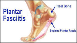
A painful and stiff sensation, sometimes compared to a sharp or stabbing pain, can be felt in the base of the heel when one is not moving around much. This condition is known as plantar fasciitis. A typical foot problem, it arises when the plantar fascia—a broad band of tissue that runs the length of the foot and provides arch support—becomes inflamed.

What are the main symptoms of plantar fasciitis?

  • Heel pain: Usually sharp and felt at the bottom of the heel, especially with the first steps in the morning or after periods of rest.
  • Pain after activity: It often worsens after exercise rather than during.
  • Stiffness: There is stiffness and discomfort in the arch of the foot. 
  • Less flexible: The foot may feel tight or less flexible, especially after long periods of standing or sitting.
  • Tenderness: Pressing on the bottom of the heel may cause discomfort.
  • Pain that improves with movement: While initial steps are painful, walking around can sometimes ease symptoms temporarily.

What causes plantar fasciitis?

Excessive strain on the plantar fascia results in plantar fasciitis, which causes irritation and inflammation. Some common causes include

  • Overuse or repetitive stress: There is overuse or excessive strain on the plantar fascia.  Activities like running, jumping, or standing for long periods put stress on the plantar fascia.
  • Foot structure: Having flat feet, high arches, or an abnormal gait can contribute to uneven pressure on the foot.
  • Improper footwear: Wearing shoes with poor arch support or thin soles can strain the plantar fascia.
  • Tight calf muscles or Achilles tendon: Limited flexibility in the lower leg can increase stress on the heel.
  • Excess body weight: Extra weight puts additional strain on the foot’s structures.
  • Sudden increase in activity: Rapidly increasing exercise intensity without proper conditioning.

What are the common treatment options for plantar fasciitis?

Treatment:

  • Rest and avoid activities that worsen the pain.
  • Stretching exercises for the foot and calf.
  • Ice therapy to reduce inflammation.
  • Supportive footwear or orthotic inserts.
  • In severe cases, doctors may recommend medical treatments like physical therapy or injections.

There are several effective treatment options for plantar fasciitis, depending on the severity of the condition. 

Here are some common approaches:

  • Home Remedies & Lifestyle Changes
  • Rest: Avoid activities that worsen the pain.
  • Ice Therapy: Apply ice packs to reduce inflammation.
  • Stretching Exercises: Stretch the plantar fascia and calf muscles to improve flexibility.
  • Supportive Footwear: Wear shoes with good arch support and cushioning.
  • Orthotic Inserts: Custom or over-the-counter insoles can help relieve pressure.

Medical Treatments


  • Nonsteroidal Anti-Inflammatory Drugs (NSAIDs): Medications like ibuprofen or naproxen help reduce pain and inflammation.
  • Physical Therapy: Strengthening and stretching exercises guided by a therapist can improve foot mechanics.
  • Corticosteroid Injections: In severe cases, doctors may recommend injections with steroids to reduce inflammation.
  • Shockwave Therapy: High-energy sound waves stimulate healing in the plantar fascia.
  • Advanced Procedures
  • Tenex Procedure: A minimally invasive technique that removes damaged tissue.
  • Surgery: Rarely needed, but in extreme cases, the plantar fascia may be surgically released from the heel bone.

Which treatments are most effective for plantar fasciitis?

The most effective treatments for plantar fasciitis depend on the severity of the condition, but here are some widely recommended options:

Highly Effective Treatments
  • Stretching & Physical Therapy: Exercises that target the plantar fascia and calf muscles can significantly reduce pain and improve flexibility.
  • Supportive Footwear & Orthotics: Wearing shoes with proper arch support and cushioning helps relieve strain on the plantar fascia.
  • Night Splints: These keep the foot in a stretched position overnight, reducing morning pain.
  • Ice Therapy: Applying ice to the affected area helps reduce inflammation and pain.
  • NSAIDs (Nonsteroidal Anti-Inflammatory Drugs): Medications like ibuprofen or naproxen can help manage pain and inflammation.

Advanced Treatments
  • Shockwave Therapy: High-energy sound waves stimulate healing in the plantar fascia.
  • Corticosteroid Injections: Used in severe cases to reduce inflammation.
  • Tenex Procedure: A minimally invasive technique that removes damaged tissue.
  • Surgery: Rarely needed, but in extreme cases, the plantar fascia may be surgically released from the heel bone.
Most people experience relief within three to six months with consistent treatment.

Explain more about physical therapy exercises.

Physical therapy exercises for plantar fasciitis focus on stretching, strengthening, and relieving tension in the foot and calf muscles. Here are some effective exercises:

Stretching Exercises
  • Towel Stretch—Sit with your legs extended, loop a towel around your foot, and gently pull it toward you.
  • Plantar Fascia Stretch—Cross one leg over the other and pull your toes back to stretch the bottom of your foot.
  • Wall Calf Stretch—Stand facing a wall, place one foot behind the other, and lean forward to stretch the calf.
Strengthening Exercises
  • Toe Curls with a Towel—Place a towel on the floor and use your toes to scrunch it up.
  • Heel Drop Stretch—Stand on a step with your heels hanging off the edge, then lower them slowly.
  • Calf Raises—Stand and lift your heels off the ground to strengthen the calf muscles.
Massage & Mobility
  • Foam Roller for the Calf—Roll a foam roller under your calf to relieve tightness.
  • Frozen Water Bottle Rolls—Roll a frozen water bottle under your foot to reduce inflammation.
  • Ball Massage—Use a tennis or lacrosse ball to massage the bottom of your foot.
These exercises help improve flexibility, reduce pain, and support healing. 

Also, read https://www.summithealth.com/plantar-fasciitis.

How Taping for Plantar Fasciitis Helps Manage Symptoms

Taping is a useful technique for managing plantar fasciitis symptoms by providing support to the foot and reducing strain on the plantar fascia. Here’s how it helps:

Benefits of Taping
  • Reduces Pain: Helps relieve discomfort by limiting excessive movement.
  • Supports the Arch: Provides stability to the foot, preventing overstretching of the plantar fascia.
  • Improves Foot Mechanics: Encourages proper alignment and reduces stress on the heel.
  • Short-Term Relief: Studies suggest taping can help manage pain temporarily.
Types of Tape Used
  • Kinesiology Tape (KT Tape): Flexible and allows movement while providing support.
  • Low-Dye Tape: Stiffer and offers more rigid support for the arch.

How to Apply Taping

  • Clean & Dry the Foot: Ensure the skin is free of moisture for better adhesion.
  • Anchor the Tape: Start at the ball of the foot and wrap around the arch.
  • Apply Strips: Use overlapping strips to reinforce support.
  • Secure the Tape: Make sure it’s snug but not too tight to restrict circulation.
Taping is often combined with stretching exercises, orthotic inserts, and physical therapy for better results. 

What Is Surgery for Plantar Fasciitis?

Surgery for plantar fasciitis is typically considered only when conservative treatments fail after 6 to 12 months of therapy. The goal is to reduce tension in the plantar fascia and relieve chronic heel pain.

Types of Surgery
  • Plantar Fascia Release—The most common procedure, where the plantar fascia is partially cut to reduce strain.
  • Endoscopic Surgery—A minimally invasive approach using small incisions and a camera for precision.
  • Open Surgery—A traditional method with a larger incision, usually reserved for severe cases.
  • Heel Spur Removal—If bone spurs are present, they may be removed during surgery.
Recovery & Risks
  • Recovery time varies from 4 to 10 weeks, depending on the procedure.
  • Risks include nerve damage, infection, and altered foot mechanics.
  • Physical therapy is often recommended post-surgery to regain strength and flexibility.
  • Most people do not require surgery, as 90% of cases improve with non-surgical treatments. 

Conclusion

Plantar fasciitis is a common yet painful condition, often caused by overuse, improper footwear, or foot structure issues. While the majority of cases improve with conservative treatments, such as stretching, supportive footwear, and physical therapy, advanced options like shockwave therapy or surgery may be considered for severe cases.

Managing plantar fasciitis requires a combination of treatment approaches, including lifestyle changes, exercises, and medical interventions. Most individuals experience significant relief within months if they follow a consistent recovery plan.

How to stop bleeding in a hemophilia patient

How to stop bleeding in a hemophilia patient

 A hemophilia overview

*Hemophilia is a hereditary condition that affects blood clotting. The condition can cause sudden or excessive bleeding, according to the CDC.

*Blood proteins called clotting factors halt bleeding. Because hemophilia patients have reduced clotting factor levels, bleeding is more likely.

*Later-life Hemophilia mainly occurs in middle-aged or older individuals or women who have given birth or are in their final months of pregnancy.

*About 1 in 5,000 male births have hemophilia. It might be mild, moderate, or severe depending on the blood clotting factor.

hemophilia
Bleeding nose hemophilia

Hemophilia types

  • Deficiency of clotting factor VIII causes hemophilia A.
  • Deficiency of clotting factor IX causes hemophilia B.
  • Acquired Hemophilia: A Rare Clotting Factor Antibody Disease.

Symptoms

  • Symptoms of hemophilia vary by severity. Some common signs:
  • There is extended bleeding from cuts, injuries, or surgery.
  • Recurring nosebleeds: Unknown causes.
  • Large, deep bruises easily appear.
  • Joint pain and swelling: Internal bleeding causes stiffness.
  • Blood in urine or stool: Internal bleeding.
  • Dental or vaccination hemorrhage: Unusual bleeding after simple operations.

Symptoms Severe

  • Unexpected bleeding.
  • Brain bleeding: Rare but serious, producing headaches, vomiting, and seizures.
  • Untreated, prolonged bleeding after surgery or injury can be fatal.

Genetics of hemophilia

  • Mutations in clotting factor genes cause hemophilia. 
  • Mutations that impair blood clotting cause profuse bleeding.

Genealogy

  • X-linked recessive hemophilia has a faulty gene on the X chromosome.
  • Males (XY) are more affected because they inherit one X chromosome. 
  • Mutations in the X chromosome cause hemophilia.
  • Carrier females (XX) have one normal and one mutant X chromosome. 
  • They may have moderate hemophilia, but it rarely gets worse.

Gene Mutations

  • Hemophilia A: Mutations in the F8 gene encode clotting factor VIII.
  • Hemophilia B: From F9 gene mutations, which encode clotting factor IX.
  • Deletions, insertions, inversions, or point mutations can alter the production or function of clotting factors.

Unplanned Changes

  • In 30% of hemophilia cases, spontaneous mutations cause the condition without a familial history.

Causes

Hemophilia is mainly hereditary. However, immune system abnormalities can cause acquired hemophilia later in life.

A genetic cause

  • Mutation: Many people inherit hemophilia. It is an X-linked recessive condition that mostly affects men but can affect women.
  • Deficiencies in clotting factors VIII and IX, respectively, cause hemophilia A and B.

Obtained Hemophilia

  • Occasionally, an autoimmune reaction that assaults clotting factors might cause hemophilia later in life.

Unplanned Mutation

  • Gene mutations can cause hemophilia without a family history.

How is hemophilia diagnosed?

 Hemophilia is diagnosed by screening and clotting factor tests to establish severity and type.

 Screening Tests
  •  These tests verify blood clotting:
  •  CBC: Measures hemoglobin and platelets.
  •  APTT Test: Measures blood clotting time.
  •  Prothrombin Time (PT) Test: Detects abnormal coagulation.
 Tests for clotting
  •  Factor Assays: Check clotting factors VIII and IX for hemophilia type and severity.
  •  Hemophilia families undergo genetic testing to identify carriers and determine risks.
 Newborn diagnosis
  •  Umbilical cord blood tests can detect hemophilia in newborns.
  •  Newborns who have significant bleeding after circumcision may undergo clotting factor tests.

 Hemophilia treatment

The video about the treatment for hemophilia


 Hemophilia treatment replaces the lacking clotting factors and stops bleeding. Some common methods:

 Standard Treatments
  •  For hemophilia A and B, factor VIII or factor IX infusions assist blood clotting.
  •  In mild hemophilia, desmopressin (DDAVP) increases coagulation.  A case.
  •  Emicizumab (Hemlibra): A novel hemophilia bleeding prevention drug.
  •  Antifibrinolytics: Clot-slowing drugs.
 Advanced Treatments
  •  Researchers are studying gene therapy as a long-term treatment for genetic defects.
  •  Non-factor therapies balance the body's natural coagulation systems without utilizing clotting factors.
  •  Supportive Care: Physical Therapy: Manages joint damage from internal bleeding.
 Lifestyle changes: avoiding aspirin and ibuprofen, and swimming or riding safely.

Furthermore, read https://www.bannerhealth.com/services/hematology/hemophilia.

 What are the patient implications of a hemophilia diagnosis?

 Hemophilia is a lifelong bleeding disease caused by a clotting factor deficiency. Thus, it implies

 Medical Effects
  •  Frequent Bleeding: Minor injuries might cause prolonged bleeding.
  •  Internal bleeding in joints can cause discomfort and movement difficulties.
  •  Cuts, surgeries, and trauma require skilled medical management to prevent severe bleeding.

 Changes in Lifestyle

  •  Avoiding Certain Drugs:  Aspirin can increase bleeding.
  •  We discourage contact sports, but swimming and cycling are safe.
  •  Patients often need clotting factor injections or novel medications.
  •  Emotional and social impact: 
  •  Managing Anxiety. Bleeding anxiety can impact daily activities.
  •  Hemophilia support groups help many patients.
  •  Career Options: Not all occupations that require physical exertion are suitable.
  •  Modern medicines allow individuals to live active, productive lives despite these limitations. 

 Genetic Testing & Progress

  •  Genetic screening detects carriers and assesses future risks.
  •  Gene therapy may be a long-term treatment for repairing defective genes.

Commonness of hemophilia
  •  Hemophilia, a rare genetic bleeding illness, differs by population.

 Global Prevalence

  •  Worldwide, 1 in 5,000 to 10,000 men have hemophilia A.
  •  Only 1 in 20,000 to 34,500 males worldwide have hemophilia B.

 The US prevalence

  •  Hemophilia affects 33,000 U.S. men.
  •  About 400 male newborns are born with hemophilia A annually.
  •  Hemophilia A is 3-4 times as frequent as B.

 Female Hemophilia

  •  Hemophilia, an X-linked recessive condition, mostly affects men and carrier women.
  •  However, some hemophiliac women have minimal bleeding.

 Hemophilia: dominant or recessive?

  •  The condition is known as X-linked recessive hemophilia. This suggests that the X chromosome is the recessive source of hemophilia.
 Why Recessive Hemophilia?
  • XY males: Since males have one X chromosome, inheriting the defective gene causes hemophilia.
  • To acquire hemophilia, females (XX) need two copies of the defective gene because they have two X chromosomes. Those who inherit one are carriers and may have modest symptoms.
 Genealogy

  •  Not dominant, hemophilia does not appear in females with one gene copy.
  •  Males cannot carry hemophilia since they have one X chromosome.

 Cure hemophilia?

 Although hemophilia is incurable, therapeutic advances have improved quality of life.

 Current Treatments

  •  Clotting Factor Replacement Therapy: Regular clotting factor supplements reduce bleeding.
 Gene Therapy: 

  • Researchers are investigating gene therapy to fix the genetic flaw for a long-term treatment.
  •  Emicizumab (Hemlibra), a new hemophilia A drug prevents bleeding.

 Future Prospects

  •  There are promising gene therapy experiments that have reduced bleeding episodes over time.
  •  Scientists create more effective and accessible remedies.
  •  Although hemophilia is lifelong, new treatments allow sufferers to live active, full lives. 

 How to stop hemophilia bleeding?

 To avoid complications, hemophilia patients must stop bleeding quickly and medically.  These stages are crucial:

 Instant Actions

  •  Press: Firmly press the bleeding location using a clean cloth or bandage.
  •  If possible, elevate the damaged limb to halt bleeding.
  •  Ice reduces edema and slows bleeding.

 Medical Care

  •  The goal is to restore clotting capacity using factor VIII or factor IX infusions for hemophilia A or B.
  •  Desmopressin (DDAVP) boosts clotting factor in mild hemophilia A.
  •  Tranexamic acid and other antifibrinolytics slow clot breakdown.
  •  To manage internal bleeding, rest and immobilize it to avoid further movement.
  •  Pain Management: Acetaminophen should replace aspirin and ibuprofen, which increase bleeding.
  •  Seek Emergency Care: Severe or internal bleeding (brain or joints) requires emergency medical attention.

Prevention of hemophilia

Hemophilia results from a defective gene, which is unpreventable. However, we can avoid complications and manage the disease.

Bleeding Prevention Methods

  • Regular clotting factor infusions reduce bleeding.
  • Avoid blood thinners: Aspirin and ibuprofen increase bleeding.
  • Protection: Helmets and padding lessen injury risk while exercising.
  • Safe Exercise: Swimming and cycling are safer than contact sports.
  • Dentists should learn about hemophilia to prevent excessive bleeding.

Genetic Advice

  • Genetic testing can identify carriers and assess future risks in hemophilia families.
  • Hemophilia-prone families may benefit from prenatal testing or genetic counseling.

Future Prospects

  • Gene Therapy Research: Scientists are investigating strategies to fix the genetic mutation and avoid hemophilia in future generations.
  • Despite the inability to prevent hemophilia, modern medicines enable individuals to lead active, fulfilling lives. 

Conclusion

Hemophilia is a lifelong inherited bleeding disorder caused by clotting factor deficits. While clotting factor replacement therapy, gene therapy research, and non-factor therapies have enhanced patients' quality of life, they cannot cure hemophilia. Hemophiliacs can live active, full lives with correct medical management, lifestyle changes, and supportive care.


The potential cure for sickle cell anemia

The potential cure for sickle cell anemia  

Overview

A series of genetic blood illnesses called sickle cell anemia are characterized by defective hemoglobin, the protein component in red blood cells that transports oxygen from the lungs to body tissues. It is the worst sickle cell illness. 

Normal hemoglobin red blood cells are disc-shaped and flexible, so they fit through large and small blood veins. In sickle cell anemia, a faulty hemoglobin S gene causes stiff protein strands in red blood cells. This shapes the cells like a sickle, a farming tool with a curved blade, giving the disease its name.

More common in Africa

Sickle-cell trait protects against malaria, a sub-Saharan African pathogen; hence, anemia and sickle-cell trait are more common in Africa. It is believed that the sickle shape makes it harder for the malaria virus to infect and easier for the body to identify and destroy infected cells. 

Causing Inflammation

These sickle-shaped cells are rigid and cling to vessel walls, creating inflammation and blood channel obstructions that restrict blood flow. Because sickle-shaped cells cannot change shape readily, they burst apart and live just 10 to 20 days, unlike normal blood cells that last three to four months.

Because it constantly makes new red blood cells to replace old ones, the body may struggle to keep up with cell destruction. This causes red blood cell deficiency and anemia. Besides anemia, the condition can cause infections, stroke, and renal damage. Long-term health difficulties can shorten life.

Sickle cell anemia

Symptoms

  • Anemia: A shortage of red blood cells causes fatigue.
  • Pain crises: Severe pain episodes brought on by obstructed blood flow.
  • Swelling: Hands and feet may swell due to circulation issues.
  • Recurrent infections: Damage to the spleen may occur.
  • Delayed growth: A lack of oxygen can slow development and puberty.
  • Vision problems: Blocked blood vessels can affect eyesight.

Causes

  • Sickle cell anemia is caused by a genetic mutation affecting hemoglobin, the protein in red blood cells that carries oxygen. A person must inherit the defective gene from both parents to develop the disease.

Treatment

Although there is no cure for everything, treatments aim to control symptoms:

  • Medications: Pain relievers, hydroxyurea (to reduce pain episodes), and L-glutamine (to minimize complications).
  • Blood transfusions: Help increase healthy red blood cells.
  • Bone marrow transplant: A potential cure, but not widely available.
  • Self-care: staying hydrated, avoiding extreme temperatures, and regular medical checkups.

What are the common complications of sickle cell anemia?

Sickle cell anemia can lead to several complications, some of which can be severe. Here are some common ones:

  • Pain crises: Episodes of intense pain due to blocked blood flow.
  • Stroke: Blood flow to the brain may be impeded by sickled cells.
  • Organ damage: Reduced oxygen supply can harm organs like the liver, kidneys, and heart.
  • Pulmonary hypertension: Breathing problems caused by elevated blood pressure in the lungs.
  • Leg ulcers: Insufficient blood flow can result in excruciating leg sores.
  • Vision problems: Blocked blood vessels can damage the retina.
  • Infections: A weakened spleen makes individuals more prone to infections.
  • Delayed growth: Oxygen deprivation can slow development in children.

Which complications are considered the most severe?

Some of the most severe complications of sickle cell anemia include:

  • Chest pain, breathing difficulties, and fever are symptoms of acute chest syndrome, a potentially fatal illness in which sickled cells obstruct blood flow to the lungs. It requires immediate medical attention.
  • Stroke: When blood supply to the brain is cut off, it can result in a stroke, which can impair speech, cause paralysis, and affect cognitive function.
  • High blood pressure in the lungs that causes heart strain and trouble breathing is known as pulmonary hypertension.
  • Organ Damage: Reduced oxygen supply can cause long-term damage to organs like the liver, kidneys, and spleen.
  • Blood Clots: Increased risk of deep vein thrombosis (DVT) and pulmonary embolism (PE), which can be fatal.

These complications require careful management and medical intervention. 

What are the treatment options for these complications?

The video explains the treatment for sickle cell anemia


Managing severe complications of sickle cell anemia requires a combination of medical treatments and lifestyle adjustments. Here are some key approaches:

  • Acute Chest Syndrome: Treated with oxygen therapy, antibiotics, blood transfusions, and pain management.
  • Stroke: Blood transfusions help reduce the risk of recurrent strokes, and some patients may benefit from hydroxyurea or bone marrow transplants.
  • Pulmonary Hypertension: Managed with medications to lower blood pressure, oxygen therapy, and regular monitoring.
  • Organ Damage: Supportive care includes medications to protect kidney function, liver health, and heart performance.
  • Blood Clots: Anticoagulants (blood thinners) help prevent dangerous clot formation.

What are the latest advancements in gene therapy for sickle cell disease?

  • Gene therapy for sickle cell disease has seen exciting advancements recently, particularly with CRISPR-based treatments. 
  • The UK and the US FDA have approved a CRISPR gene therapy for sickle cell anemia and beta thalassemia, marking a major milestone in genetic medicine.
  • Additionally, several clinical trials are underway
  • Gene therapy's looking promising for sickle cell, especially with CRISPR offering hope for a potential cure.

How Is Sickle Cell Anemia Diagnosed?

Sickle cell anemia is diagnosed through various tests that check for the presence of abnormal hemoglobin. Here are the main methods:

  • Blood Tests: A blood sample is analyzed to detect sickle-shaped red blood cells and abnormal hemoglobin.
  • Hemoglobin Electrophoresis: This test identifies different types of hemoglobin, including hemoglobin S, which causes sickle cell disease.
  • Complete Blood Count (CBC): Measures red blood cell levels and checks for anemia.
  • Peripheral Blood Smear: Examines the shape of red blood cells under a microscope.
  • Genetic Testing: Determines if a person carries the sickle cell gene.
  • Prenatal Screening: Amniotic fluid or placental tissue is tested to diagnose sickle cell anemia in unborn babies.
  • Newborn Screening: A heel-prick blood test is performed on newborns to detect sickle cell disease early.
Also, read https://www.pfizer.com/disease-and-conditions/sickle-cell.

Prognosis of Sickle Cell Anemia

Over time, improvements in medical care and treatment options have led to a significant improvement in the prognosis for sickle cell anemia. While it remains a lifelong condition, many individuals with sickle cell anemia can live into their 50s or beyond with proper management.

Factors Affecting Prognosis:

  • Life Expectancy: Historically, life expectancy was lower, but improved treatments have extended survival rates.
  • Complications: Severe complications like stroke, acute chest syndrome, and organ damage can impact prognosis.
  • Medical Advances: Hydroxyurea, blood transfusions, and emerging gene therapies are improving outcomes.
  • Preventive Care: Regular checkups, vaccinations, and lifestyle adjustments help manage symptoms and reduce complications.

Duration of Sickle Cell Anemia

  • Sickle cell anemia is a lifelong condition, meaning it does not go away over time. However, with advancements in treatment, individuals with sickle cell anemia can live into their 50s or beyond.
  • The duration of symptoms and complications varies depending on the severity of the disease and the effectiveness of treatment. Some people experience frequent pain crises and complications, while others have milder symptoms.

Is it possible to prevent sickle cell disease?

Being a genetic disorder that is inherited, sickle cell anemia cannot be totally prevented. However, there are ways to reduce the risk of passing it on and manage its complications effectively.

Prevention Strategies:

  • Genetic Counseling: Speaking with a genetic counselor can help determine your child's risk of inheriting the sickle cell trait if you or your partner has it.
  • Prenatal Screening: Testing during pregnancy can determine if the baby has sickle cell disease.
  • Newborn Screening: Early detection allows for better management and preventive care.
  • Lifestyle Adjustments: While not preventing the disease itself, staying hydrated, avoiding extreme temperatures, and getting regular medical checkups can help prevent complications.

Stem cell or bone marrow transplants for sickle cell disease

Stem cell or bone marrow transplants are currently the only potential cure for sickle cell disease. These transplants replace the defective blood-forming cells with healthy ones from a donor, allowing the body to produce normal red blood cells.

How It Works:

  • Finding a Donor: A matched sibling or unrelated donor is needed.
  • Preparation: Patients undergo chemotherapy to weaken their immune system and prevent rejection.
  • Transplant Procedure: Healthy stem cells are infused into the bloodstream, where they travel to the bone marrow and start producing normal blood cells.
  • Recovery: It can take 6-12 months for the immune system to rebuild.

Risks and Challenges:

  • Finding a Match: Only 20 to 30% of children have a sibling match.
  • Rejection & Complications: The body may reject the new cells, leading to graft-versus-host disease.
  • Long Recovery: Patients require months of monitoring to ensure success.

Latest Advancements:

  • Reduced-intensity transplants: New approaches minimize the need for strong immunosuppressing drugs.
  • Expanded donor options: Researchers are exploring half-matched family donors.
  • Gene therapy: Emerging treatments aim to correct the genetic mutation without needing a donor.

Conclusion

Sickle cell illness was life-threatening decades ago. Recently improved treatments have extended and enhanced the quality of life for those with the illness.  

People with sickle cell disease may live to 50 or older, but not as long as others. Death often results from organ failure or infections.


Animals spread deadly bubonic plague

Animals spread the deadly bubonic plague

Overview

In humans, three types of plague have been identified. Most cases of plague are caused by flea bites from infected animals; this type is known as bubonic plague (Poland and Barnes, 1979). Lymph nodes that drain from the site of infection become inflamed and sensitive, eventually creating a bubo as the bacteria spread from there. It is also called the Black Death. The hallmark of bubonic plague is the bubo, which can grow to be as big as an egg. Blood culture numbers below 10^-4×10⁷ cfu/ml have the potential to cause bacteremia, according to Perry and Fetherston (1997).


bubonic plague
BUBO BACTERIA

Key Symptoms of Bubonic Plague:

Bubonic plague is a serious bacterial infection caused by Yersinia pestis, primarily transmitted through flea bites. Here are the key symptoms:

*Fever and chills—A sudden high fever is common.

*The patient is experiencing headache and weakness, general fatigue, and discomfort.

*Swollen lymph nodes (buboes) are painful, enlarged lymph nodes that are typically located in the groin, armpit, or neck.

*Muscle aches refer to general body pain and soreness.

*Skin changes—In severe cases, skin may darken due to tissue death.

If untreated, the infection can spread and lead to septicemic or pneumonic plague, which are even more dangerous. If you notice any symptoms, please seek medical attention promptly.

What caused the bubonic plague

  • The bubonic plague is caused by the bacterium Yersinia pestis, which primarily spreads through fleas that infest small mammals like rats. When an infected flea bites a human, the bacteria enter the body and travel through the lymphatic system, leading to swollen lymph nodes (buboes), fever, and other symptoms.
  • Historically, the plague has been responsible for devastating pandemics, including the Black Death in the 14th century, which wiped out millions across Europe, Asia, and Africa. The disease can also spread through direct contact with infected animals or their bodily fluids.

Bubonic plague treatment

Bubonic plague is treatable with antibiotics if caught early. Common treatments include:

*Antibiotics—Medications like ciprofloxacin, levofloxacin, moxifloxacin, gentamicin, and doxycycline are effective.

*Supportive care—Patients may need fluids, oxygen, and medications to manage symptoms.

*Isolation—To stop the spread of the infection, isolating individuals may be necessary.

*Preventive measures—If exposed, prophylactic antibiotics can help prevent infection.

*Early diagnosis and treatment are critical to survival. 

Also read https://nypost.com/article/what-is-bubonic-plague/.

How did the bubonic plague spread

*The bubonic plague spread primarily through fleas that carried the bacterium Yersinia pestis. 

*These fleas infested rodents, such as rats, which traveled along trade routes and into human settlements.

*When infected fleas bite humans, the bacteria enter the body, leading to infection.

*The plague also spread through human-to-human transmission, especially in its pneumonic form, which affected the lungs and could be passed through respiratory droplets. 

*In medieval times, poor sanitation and crowded living conditions accelerated its spread.

What were the social impacts of the bubonic plague?

The bubonic plague had profound social impacts, reshaping societies in ways that lasted for centuries. Here are some key effects:

Breakdown of Social Order—Fear and death led to widespread panic. Many abandoned their families, fled cities, or isolated themselves.

Religious Upheaval—Faith in the Church declined as prayers failed to stop the plague. Some turned to extreme religious movements, while others lost trust in religious institutions.

Economic Shifts: A massive population decline led to a shortage of labor. Peasants gained more bargaining power, leading to higher wages and better working conditions.

Rise of Social Mobility—The rigid feudal system weakened as serfs moved freely to find better opportunities. This contributed to the decline of feudalism.

Persecution and Scapegoating—Minority groups, especially Jews, were falsely blamed for the plague and faced violent persecution.

Cultural Changes—Art and literature reflected themes of death and despair. The plague influenced a shift toward more individualistic and humanistic perspectives, paving the way for the Renaissance.

The plague didn’t just devastate populations—it transformed medieval society. 

How did different governments respond to the plague?

Governments throughout history responded to the bubonic plague in various ways, often shaped by their understanding of disease and available resources. Here are some key approaches:

Quarantines & Isolation—Cities like Venice pioneered quarantine measures, requiring ships to wait 40 days before docking. This practice later became standard across Europe.

Public Health Regulations—Some governments imposed sanitation laws, banning gatherings and improving waste disposal to limit the spread.

Travel Restrictions: To stop infected people from spreading the disease, we closed borders and restricted movement between towns.

Religious & Superstitious Responses—Many rulers turned to prayers, processions, and spiritual rituals, believing the plague was divine punishment.

Economic Adjustments—With labor shortages, governments introduced wage controls and laws to stabilize economies.

Medical Advancements—The repeated outbreaks led to early public health boards and medical studies that shaped modern epidemiology.

Despite these efforts, the plague often overwhelmed societies, forcing rulers to adapt their strategies over time. 

How did the Black Plague end? (Watch the video.)


The Black Plague gradually declined due to several factors:

Improved Hygiene & Sanitation—As people learned more about disease transmission, cities began implementing better sanitation practices.

Quarantines & Isolation—Many regions enforced strict quarantines, preventing infected individuals from spreading the disease.

Natural Immunity—Some survivors developed immunity, reducing the number of susceptible hosts.

Shifts in climate and a decline in infected rodent populations slowed the spread.

Medical Advancements—Over time, better medical knowledge and treatments have helped control outbreaks.

Although the Black Plague faded, bubonic plague continued to appear in smaller outbreaks for centuries. Today, it is treatable with antibiotics if caught early.

How long did the Black Plague last

The Black Plague, also known as the Black Death, lasted from 1346 to 1353. It was one of the deadliest pandemics in history, killing an estimated 25 to 50 million people across Europe, Asia, and North Africa. While the initial outbreak lasted about seven years, smaller outbreaks continued for centuries, with recurrences well into the 19th century.

Was the Black Death 100% fatal?

No, the Black Death was not 100% fatal, but it had an extremely high mortality rate. The survival chances depended on the type of plague:

The most common form of the plague, known as bubonic plague, has a fatality rate of 30-75% if left untreated. While some people recovered, many suffered lasting effects.

The pneumonic plague is a more severe form that affects the lungs, with a near 100% fatality rate if left untreated.

The septicemic plague is the deadliest form, causing rapid bloodstream infection and having a near 100% fatality rate.

Overall, the Black Death wiped out 30–60% of Europe's population, but some individuals survived, likely due to genetic resistance or immunity.

Conclusion

Although bubonic plague seems ancient, it still thrives in rural America and around the world.  Avoiding fleas on rats, mice, and squirrels is the greatest way to be free from plague.  Bunnies and chipmunks can also have fleas.  Protect your pets and yourself from fleas and their diseases.  Contact your doctor if you have a high fever, chills, or swollen, painful lymph nodes.


Drugs that cause tinnitus (ringing ears)

Drugs that cause tinnitus (ringing ears) 

Describe Tinnitus

Tinnitus is a condition where a person hears ringing, buzzing, clicking, or other phantom sounds in one or both ears, even when there is no external source of the noise. About 15% to 20% of people suffer from it, primarily older adults. A few drugs can cause tinnitus.

tinnitus (ringing ears)
Drugs may cause tinnitus 

Symptoms

  • People with tinnitus may experience:
  • Ringing, buzzing, clicking, or hissing sounds.
  • Sounds that vary in pitch and intensity.
  • Phantom noises can be either constant or intermittent.

Types of tinnitus

Tinnitus comes in different forms, each with unique characteristics and causes. Here are the main types:

1. Subjective Tinnitus

  • The most prevalent type.
  • Only the affected person can hear the ringing or buzzing.
  • Exposure to loud noise, ear infections, or certain medications can cause ear infections.

2. Objective Tinnitus

  • An uncommon variety in which a physician can hear the sound through a stethoscope.
  • It is usually linked to vascular or muscular issues, such as aneurysms or muscle spasms.

3. Somatic Tinnitus

  • The intensity of the sound changes with body movement.
  • This condition is often associated with issues in the neck or jaw, such as temporomandibular joint (TMJ) disorders.

4. Pulsatile Tinnitus

  • There is a rhythmic noise that matches the heartbeat.
  • This condition is often caused by changes in blood flow near the ear.

5. Musical Tinnitus (Musical Hallucination)

  • This refers to the perception of music or singing when no music is playing.
  • This condition is more common in people with hearing loss or neurological conditions.
  • Each type has different treatment approaches, depending on the underlying cause. 

Tinnitus's origins

Although tinnitus can be caused by a variety of factors, it is often linked to issues with the ears or nervous system. Here are some common causes:

1. Hearing Loss

  • Tinnitus results from age-related hearing loss, which is caused by deterioration of the inner ear.
  • Noise-induced hearing loss— Exposure to loud sounds can damage the ear’s hair cells.

2. Ear Conditions

  • Earwax accumulation: Too much earwax can obstruct the ear canal and result in tinnitus.
  • Ear infections—Inflammation or fluid buildup can trigger ringing in the ears.
  • Ménière’s disease—A disorder affecting the inner ear, often accompanied by dizziness.

3. Circulatory System Issues

  • High blood pressure—Increased blood flow near the ear can cause pulsatile tinnitus.
  • Atherosclerosis—Narrowed arteries can lead to abnormal blood flow sounds.
  • Vascular disorders—Conditions affecting blood vessels near the ear may contribute.

4. Neurological & Musculoskeletal Causes

  • Temporomandibular joint (TMJ) disorders—jaw problems can affect ear function.
  • Head or neck injuries—Trauma can disrupt auditory pathways.
  • Overactive thyroid—Hormonal imbalances may contribute to tinnitus.

5. Medications & Other Factors

  • Certain drugs—some antibiotics, antidepressants, and anti-inflammatory medications—can trigger tinnitus.
  • Smoking & alcohol—These substances can affect circulation and worsen symptoms.
  • Stress & anxiety—Psychological factors can heighten tinnitus perception.

Medical conditions of tinnitus

Tinnitus can be linked to several medical conditions, often affecting the ears, nervous system, or circulatory system. Here are some common conditions associated with tinnitus:

Ear & Hearing Conditions

  • Age-related hearing loss—The inner ear deteriorates over time, leading to tinnitus.
  • Noise-induced hearing loss— Exposure to loud sounds can damage the ear’s hair cells.
  • Ménière’s disease—A disorder affecting the inner ear, often accompanied by dizziness and hearing loss.
  • Ear infections—Inflammation or fluid buildup can trigger ringing in the ears.
  • Earwax blockage—Excess earwax can obstruct the ear canal and cause tinnitus.
  • Neurological & Musculoskeletal Conditions
  • Temporomandibular joint (TMJ) disorders—jaw problems can affect ear function.
  • Head or neck injuries—Trauma can disrupt auditory pathways, leading to tinnitus.
  • Multiple sclerosis is a neurological disorder that can affect hearing and cause tinnitus.

Circulatory System Disorders

  • High blood pressure—Increased blood flow near the ear can cause pulsatile tinnitus.
  • Atherosclerosis—Narrowed arteries can lead to abnormal blood flow sounds.
  • Vascular malformations—Abnormal blood vessel structures near the ear may contribute.

Other Medical Conditions

  • Diabetes can affect circulation and nerve function, potentially leading to tinnitus.
  • Thyroid disorders—Hormonal imbalances may contribute to tinnitus symptoms.
  • Autoimmune diseases—Conditions like lupus or rheumatoid arthritis may have links to tinnitus.

Tinnitus is often a symptom rather than a standalone condition, so addressing the underlying medical issue can help manage its effects. If tinnitus is persistent or worsening, consulting a healthcare provider is recommended.

Medications that cause tinnitus

Certain medications can cause or worsen tinnitus as a side effect. These drugs are often referred to as ototoxic, meaning they can harm the inner ear. Here are some common types:

1. Pain Relievers (Analgesics)

  • High-dose aspirin
  • Ibuprofen (Advil, Motrin)
  • Naproxen (Aleve)
  • Acetaminophen (Tylenol)

2. Antibiotics

  • Aminoglycosides (e.g., gentamicin, neomycin)
  • Macrolides (e.g., erythromycin, azithromycin)

3. Chemotherapy Drugs
  • Cisplatin
  • Carboplatin
4. Diuretics (Water Pills)

  • Furosemide (Lasix)
  • Bumetanide

5. Antidepressants & Anti-Anxiety Medications

  • Selective serotonin reuptake inhibitors (SSRIs) (e.g., fluoxetine, sertraline)
  • Tricyclic antidepressants (e.g., amitriptyline, nortriptyline)
  • Benzodiazepines (e.g., diazepam, lorazepam)

6. Blood Pressure Medications

  • Beta blockers (e.g., propranolol)
  • ACE inhibitors (e.g., lisinopril)

7. Anti-Malarial Drugs

  • Chloroquine
  • Quinine

Tinnitus caused by medications is often temporary and may resolve once the drug is discontinued. However, in some cases, it can be permanent. If you suspect a medication is causing tinnitus, consult a healthcare provider before making any changes.

Also read https://www.britannica.com/science/tinnitus.

Tinnitus diagnosis

Tinnitus diagnosis typically involves a combination of medical history review, physical examination, and specialized tests to determine the underlying cause. Here’s how it’s generally assessed:

1. Medical History & Physical Examination

  • Your doctor will ask about your symptoms, including the type of sounds you hear, their frequency, and any associated conditions.
  • They may check for earwax buildup, ear infections, or jaw and neck issues that could contribute to tinnitus.

2. Hearing Tests (Audiometry)
  • You may undergo a hearing exam in a soundproof room, where specific sounds are played to assess your hearing ability.
  • This technique helps determine if tinnitus is linked to hearing loss.
3. Imaging Tests
  • CT scans or MRI scans may be recommended if structural abnormalities or tumors are suspected.
  • These tests help rule out vascular disorders, acoustic neuromas, or other neurological conditions.
4. Blood Tests
  • Blood tests may be conducted to check for anemia, thyroid disorders, or high blood pressure, which can contribute to tinnitus.
5. Movement & Neurological Tests

  • Your doctor may ask you to move your eyes, clench your jaw, or turn your neck to see if tinnitus changes.
  • This can help identify musculoskeletal causes like TMJ disorders.

6. Pulsatile Tinnitus Assessment

  • If your tinnitus follows your heartbeat, it may be pulsatile tinnitus, often linked to circulatory system issues.
  • To examine blood vessel abnormalities, additional tests might be required.

Since tinnitus can have multiple causes, a thorough diagnosis is essential for determining the best treatment approach. If you’re experiencing persistent tinnitus, consulting an ENT specialist or audiologist is recommended.

Tinnitus Treatment & Management

The video explains how to manage tinnitus


While there is no universal cure, tinnitus can be managed through

Medical Treatments

  • Treating underlying conditions—addressing issues like earwax buildup, infections, or circulatory problems.
  • Hearing aids—useful for those with tinnitus linked to hearing loss.
  • Medications—Some antidepressants and anti-anxiety drugs may help reduce tinnitus perception.

Therapies

  • Sound therapy—using white noise machines or soothing sounds to mask tinnitus.
  • Cognitive behavioral therapy (CBT)—Helps patients cope with tinnitus-related distress.
  • Tinnitus retraining therapy (TRT)—combines counseling and sound therapy to help the brain ignore tinnitus.

Lifestyle & Home Remedies

  • Avoid loud noises—protect your ears from excessive noise exposure.
  • Reduce stress—stress management techniques like meditation and deep breathing can help.
  • Limit caffeine and alcohol—these substances may worsen tinnitus symptoms.
  • Exercise and a healthy diet—maintaining overall health can improve tinnitus management.

Treatment & Management

  • Lifestyle changes—reducing caffeine intake, managing stress, and avoiding loud noises.
  • If tinnitus is persistent or interferes with daily life, consulting a healthcare provider is recommended. 

Alternative Treatments

  • Acupuncture—Some people find relief through acupuncture sessions.
  • Herbal supplements—Certain supplements like ginkgo biloba are believed to help, though scientific evidence is limited.
  • If tinnitus is persistent or worsening, consulting an ENT specialist or audiologist is recommended. You can find more details on tinnitus treatment here. 

How to prevent tinnitus

Preventing tinnitus involves protecting your ears and maintaining overall health. Here are some effective strategies:

1. Protect Your Ears

  • Avoid loud noises—prolonged exposure to loud sounds can damage hearing.
  • In noisy situations, use ear protection by donning noise-canceling headphones or earplugs.
  • Keep volume levels low—when using headphones, follow the 60/60 rule (60% volume for no more than 60 minutes at a time).

2. Maintain Good Health
  • Manage blood pressure—high blood pressure can contribute to tinnitus.
  • Exercise regularly—it improves circulation and overall ear health.
  • Eat a balanced diet—nutrients like magnesium and zinc may support ear function.
3. Reduce Risk Factors

  • Limit caffeine and alcohol—these substances can worsen tinnitus symptoms.
  • Avoid smoking—smoking affects circulation and can increase tinnitus risk.
  • Manage stress—stress and anxiety can heighten tinnitus perception.

4. Take Care of Your Ears
  • Avoid excessive earwax buildup—clean ears safely without inserting objects.
  • Treat ear infections promptly—untreated infections can lead to complications.
  • Stay hydrated—proper hydration supports inner ear function.

Natural treatments for tinnitus

Alternative treatments for tinnitus focus on natural and holistic approaches to managing symptoms. While scientific evidence varies, some people find relief through these methods:

1. Herbal Supplements

  • Ginkgo biloba—believed to improve blood circulation to the ears.
  • Acai Red Asian ginseng—May support nerve health.
  • Zinc & magnesium—essential minerals that may aid ear function.

2. Acupuncture

  • Some individuals report reduced tinnitus symptoms after acupuncture sessions.
  • Thought to help by improving circulation and reducing stress.

3. Mindfulness & Relaxation Techniques

  • Meditation & yoga can help manage stress, which may worsen tinnitus.
  • Deep breathing exercises may reduce anxiety linked to tinnitus perception.

4. Sound Therapy

  • White noise machines—mask tinnitus sounds with soothing background noise.
  • Music therapy—Listening to altered frequencies may help retrain the brain.

5. Chiropractic & TMJ Treatment

  • Chiropractic adjustments—May help if tinnitus is linked to neck or jaw issues.
  • TMJ therapy—Treating jaw disorders can reduce tinnitus symptoms.

6. Dietary Changes

  • Reducing caffeine & alcohol—These substances may worsen tinnitus.
  • Eating anti-inflammatory foods and a balanced diet may support ear health.

While these treatments may help some individuals, results vary. If tinnitus is persistent, consulting a healthcare provider is recommended. 

Conclusion

Tinnitus is a complex condition that can stem from various causes, including hearing loss, circulatory issues, and certain medications. While there is no universal cure, many treatment options—from sound therapy and cognitive behavioral therapy to lifestyle adjustments and alternative remedies—can help individuals manage symptoms effectively. Protecting your ears, maintaining good overall health, and seeking professional guidance when needed are key steps in preventing and managing tinnitus. If symptoms persist or worsen, consulting a healthcare provider is highly recommended.

Home remedies for intertrigo skin disorder.

Home remedies for intertrigo skin disorder.

Overview Intertrigo

Skin-to-skin friction causes intertrigo, a common inflammatory skin condition that is frequently made worse by heat and moisture. In places where skin folds, like the armpits, groin, under the breasts, and in between fingers or toes, it usually manifests as a red, bumpy rash.

While intertrigo itself is not an infection, it can lead to secondary bacterial or fungal infections, with Candida (yeast) being a frequent culprit. People who are overweight, diabetic, or experience excessive sweating are more prone to developing intertrigo.

Treatment usually involves keeping the affected area dry, using antifungal or antibacterial medications if an infection occurs, and applying low-potency topical steroids to reduce inflammation. Preventative measures include good hygiene, weight management, and reducing skin friction.

intertrigo
intertrigo infections


What are the symptoms of intertrigo?

Intertrigo presents as an inflamed rash in areas where skin rubs together, often worsened by heat and moisture. Here are the common symptoms:

Key Symptoms

  • Red or reddish-brown rash (may appear darker in deeper skin tones)
  • Itching, burning, or stinging sensation
  • Raw or oozing skin in severe cases
  • Foul odor if a secondary infection develops
  • Cracked or crusty skin
  • Pain or discomfort, especially in skin folds

Commonly Affected Areas

  • Armpits
  • Under the breasts
  • Groin and inner thighs
  • Between fingers and toes
  • Neck folds
  • Abdominal skin folds
  • Between the buttocks

Intertrigo itself is not an infection, but it can lead to secondary bacterial or fungal infections, often caused by Candida (yeast). 

What causes intertrigo

Heat, moisture, and lack of air circulation exacerbate skin-to-skin friction, which causes intertrigo. Here are the main contributing factors:

Primary Causes

  • Friction: Constant rubbing of skin folds leads to irritation.
  • Moisture & Sweat: Trapped moisture prevents evaporation, creating an ideal environment for inflammation.
  • Heat & Humidity: Hot climates increase sweating, making intertrigo more likely.
  • Lack of Airflow: Poor ventilation in skin folds keeps moisture trapped.

Risk Factors

  • Obesity: Excess skin folds increase friction and moisture retention.
  • Diabetes: Higher risk due to changes in skin health and immune function.
  • Excessive Sweating: Hyperhidrosis can worsen intertrigo.
  • Poor Hygiene: Unclean skin folds can lead to bacterial or fungal overgrowth.
  • Smoking & Alcohol: These can increase susceptibility to intertrigo.

Intertrigo itself is not an infection, but it can lead to secondary bacterial or fungal infections, often caused by Candida (yeast).

What are effective ways to prevent intertrigo?

The video about intertrigo treatment


Preventing intertrigo involves keeping skin folds dry, reducing friction, and maintaining excellent hygiene. Here are some effective strategies:

1. Keep Skin Dry

  • Pat dry after washing. Avoid rubbing, as it can worsen irritation.
  • Use absorbent powders: Talcum or cornstarch-based powders help absorb excess moisture.
  • Air out skin folds: Allow airflow by wearing loose clothing.

2. Reduce Friction
  • Wear breathable fabrics: cotton or moisture-wicking materials minimize irritation.
  • Apply barrier creams, such as zinc oxide or petroleum-based creams, to create a protective layer.
3. Maintain Good Hygiene

  • Clean skin folds regularly. Wash with mild soap and water, then dry thoroughly.
  • Avoid excessive sweating: Stay cool and dry, especially in humid conditions.

4. Lifestyle Adjustments

  • Maintain a healthy weight: Reducing skin folds lowers friction.
  • Stay hydrated: Proper hydration supports skin health.

What factors increase the risk of intertrigo?

Several factors can increase the risk of developing intertrigo, a skin condition caused by friction, moisture, and heat in skin folds. Here are some key risk factors:

1. Environmental Factors

  • Hot and humid climates: Increased sweating and moisture retention worsen intertrigo.
  • Poor ventilation: Lack of airflow in skin folds encourages irritation.

2. Health & Lifestyle Factors

  • Obesity: Excess skin folds lead to more friction and trapped moisture.
  • Diabetes: Higher risk due to changes in skin health and immune function.
  • Excessive sweating (hyperhidrosis) creates a moist environment ideal for irritation.
  • Poor hygiene: Unclean skin folds can lead to bacterial or fungal overgrowth.

3. Other Contributing Factors

  • Wearing tight clothing increases friction and prevents proper airflow.
  • Smoking & alcohol consumption can weaken skin health.
  • Weakened immune system: Conditions like HIV or chemotherapy can make skin more vulnerable.

How is intertrigo diagnosed?

Intertrigo is typically diagnosed through a clinical examination by a healthcare provider. Here’s how the process usually works:

1. Physical Examination

  • A doctor will inspect the affected skin folds for redness, irritation, and signs of infection.
  • They may ask about symptoms like itching, burning, or odor.

2. Diagnostic Tests (If Needed)

  • Skin swab or scraping: If an infection is suspected, a sample may be taken to check for bacteria or fungi.
  • Wood’s lamp test: A special UV light can help detect certain fungal infections.
  • KOH test: A potassium hydroxide (KOH) preparation can confirm the presence of Candida (yeast).
  • Rarely, a small skin sample may undergo a biopsy under a microscope.

3. Differential Diagnosis

  • Doctors may rule out other skin conditions like eczema, psoriasis, or allergic reactions.
  • Would you like to know how to manage intertrigo effectively? You can also check out more details here or here.

What are the most effective treatments for intertrigo?

The most effective treatments for intertrigo focus on keeping the affected area dry, reducing friction, and addressing any secondary infections. Here are some key approaches:

1. Hygiene & Prevention

  • Keep the area clean and dry: Wash gently with mild soap and water, then pat dry.
  • Wear loose, breathable clothing: cotton or moisture-wicking fabrics help reduce friction and sweating.
  • Use absorbent powders: Talcum or cornstarch-based powders can help keep the skin dry.

2. Topical Treatments

  • Antifungal creams: If a fungal infection is present, clotrimazole or miconazole creams can be effective.
  • Antibacterial ointments: Doctors may prescribe topical antibiotics like mupirocin if bacteria are present.
  • Barrier creams: Zinc oxide or petroleum-based creams create a protective layer to reduce irritation.
  • Low-potency corticosteroids: Hydrocortisone creams can help reduce inflammation and itching.

3. Medical Treatments

  • Oral antifungals or antibiotics: In severe cases, doctors may prescribe oral medications to treat infections.
  • Professional evaluation: We recommend consulting a healthcare provider if symptoms persist or worsen.

Would you like recommendations for specific products or home remedies?

Here are some home remedies and over-the-counter products that can help manage intertrigo:

Home Remedies

  • Cornstarch or Talcum Powder: Helps absorb excess moisture and reduce friction.
  • Coconut Oil: Has mild antibacterial and antifungal properties to soothe irritation.
  • Aloe Vera Gel: Provides cooling relief and supports skin healing.
  • Apple Cider Vinegar (Diluted): Acts as a natural antifungal when applied lightly.
  • Tea Tree Oil (Diluted): Known for its antibacterial and antifungal effects.

Over-the-Counter Products

  • Antifungal creams: Clotrimazole, miconazole, or terbinafine can help with yeast infections.
  • Barrier creams: Zinc oxide or petroleum-based ointments create a protective layer.
  • Hydrocortisone cream: Helps reduce itching and inflammation (use sparingly).
  • Antibacterial ointments: Mupirocin or neomycin help combat bacterial infections.

Would you like to know how to prevent it or manage flare-ups?

Prevention Tips

  • Keep skin folds dry: Pat dry after washing, and use absorbent powders.
  • Wear breathable fabrics: cotton or moisture-wicking materials help reduce moisture.
  • Apply barrier creams: zinc oxide or petroleum jelly can protect skin from friction.
  • Maintain hygiene: Regularly cleanse affected areas with mild soap.
  • Manage weight and sweat: Excess skin folds and sweating increase risk.

Managing Flare-Ups

  • Use antifungal or antibacterial creams if an infection develops.
  • Apply hydrocortisone cream to reduce inflammation (use sparingly).
  • Keep the area cool and aerated to speed healing.
  • Avoid irritants like harsh soaps that can worsen symptoms.

Conclusion

Intertrigo is a common skin condition caused by friction, moisture, and heat, often affecting areas where skin folds. While it’s not an infection, it can lead to secondary bacterial or fungal complications if untreated.

Effective management involves keeping the skin dry, using antifungal or antibacterial treatments when necessary, and reducing friction with barrier creams and breathable clothing. Preventive strategies like maintaining excellent hygiene, managing weight, and minimizing excessive sweating can significantly reduce the risk.